Imaging Core
 The Aga Khan University (Pakistan) hosts an imaging suite equipped with advanced imaging technologies and software, including:
Location
Imaging Suite
BSL-2 Laboratory, First Floor
Juma Building
Aga Khan University, Stadium Road
Access
The facilities are accessible to AKU and non-AKU users at competitive rates for
research purposes only.
Publications Policy
For publications that include data generated using equipment and software at the Imaging Core, it is mandatory to share the mansuscript with the relevant teams to ensure accurate reporting and interpretation of data.
Virtual Microscope
The virtual microscope creates digital images from routine glass slides, which can be viewed on computer monitors in a manner that simulates real observation sans the microscope. These images can be used for teaching, research, and archival use by researchers, shared by groups, and can be incorporated into teleconferences and digital pathology.
Services
-
Whole slide scanning at 2x 10x, 20x, 40x
-
Z compression at 2x 10x, 20x, 40x
-
Z-stack at 2x 10x, 20x, 40x
-
Tissue Micro-Array 2x 10x, 20x, 40x
For queries and appointments
Dr Azhar Hussain, Director
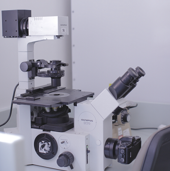
Confocal Microscope
A1R plus is a high-end point scanning confocal microscope. The system features a "hybrid scanner," a glavano scanner for high resolution up to 16 MP and a resonant scanner for ultrahigh speed imaging at 420fps (512 x 32 pixels). Using standard detector up to 4-color high resolution imaging can be performed by four lasers. Flourophore with overlapped spectra can be imaged using spectral detector.
Techniques
Four color confocal microscopy
Multipoint and live time-lapse imaging
Spectral detection and unmixing
Transmitted light Differential Interference Contrast (DIC)
Up to 6 dimensional Imaging on Live and Fixed cells
Fluorescence recovery after photobleaching (FRAP)
Fluorescence Resonance Energy Transfer (FRET)
Calcium Imaging
Photoactivation
Colocalization
Instrument Specifications
Inverted Nikon Ti E microscope
Objectives 10x, 20x 40x dry objectives. 60x & 100x oil immersion
Four lasers: L1 - a solid state 640nm, L2 - a solid state 405nm, L3 - an Argon gas laser with 457nm, 477nm, 488nm & 514nm laser lines and L4 - a solid state 561nm
Four PMT detectors
32 channel spectral detector
Perfect focus system (PFS) for time lapse imaging
Tokai Hit incubator for time lapse imaging
NIS Elements software (AVI movies for time lapse)
For queries and appointments
Fluorescence Activated Cell Sorter
Flowcytometry is the measurement of cell properties in a flowing medium. It can be used by researchers to carry outimmunophenotyping and analyse cell cycle and cellular material such as DNA and chromosomes from a range of cell types.
Orientation/Training
Orientation of the facility is mandatory for all users. Users will be briefed on the general principles of the flowcytometry and the guidelines of setting up their experiments. These sessions will be conducted by dedicated staff and faculty. The purpose of this training is to provide knowledge to users that will help them in designing their experiment efficiently and to use the facility effectively.
For queries and appointments
Abdul Wahab Siddiqui, Research Associate
---------------------------------------------------------------------------------------------------------------------------------------------
You can learn more about these equipments
Useful spectrum viewers
E-Learning (flowcytometry, software, and hardware theory
