K-VersionIt includes two simulators: Cardiology
Patient Simulator (K) and Lung Sound Auscultation
Trainer (LSAT). Simulator K has 88 examples of comprehensive simulation
of patients with heart diseases and arrhythmias whereas simulator LSAT has 36
lung sound cases both posterior and anterior. These Sounds are recorded
from actual patients and are reproduced by using a high quality sound
system.
| 
|
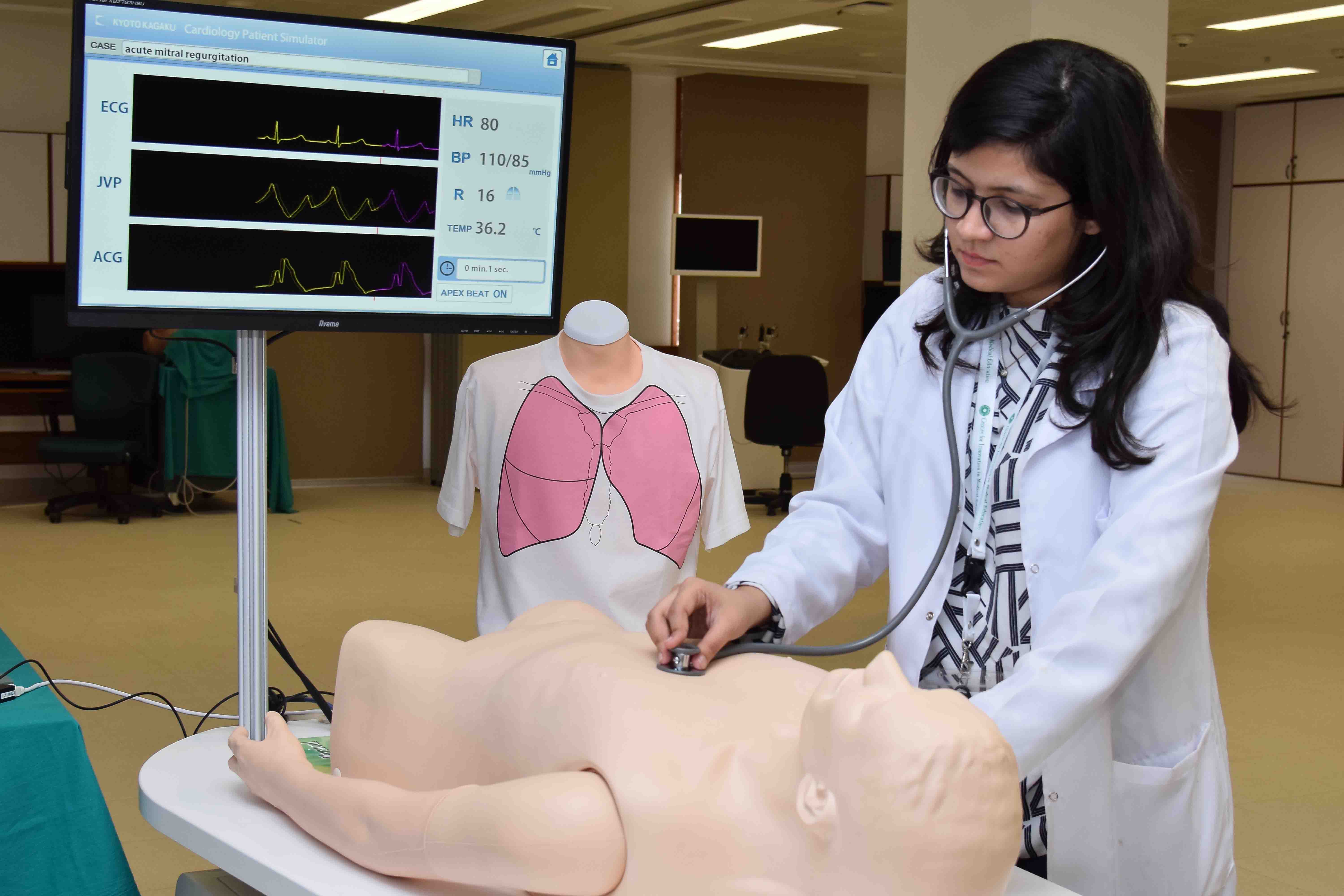
Harvey (Cardiopulmonary Patient Simulator)Harvey has been a proven simulation system to teach bedside cardiac assessment skills that transfer to real patients. Harvey, The Cardiopulmonary Patient Simulator, is the longest continuous university-based simulation project in medical education. It realistically simulates nearly any cardiac disease at the touch of a button by varying blood pressure, pulses, heart sounds, murmurs and breath sounds. | 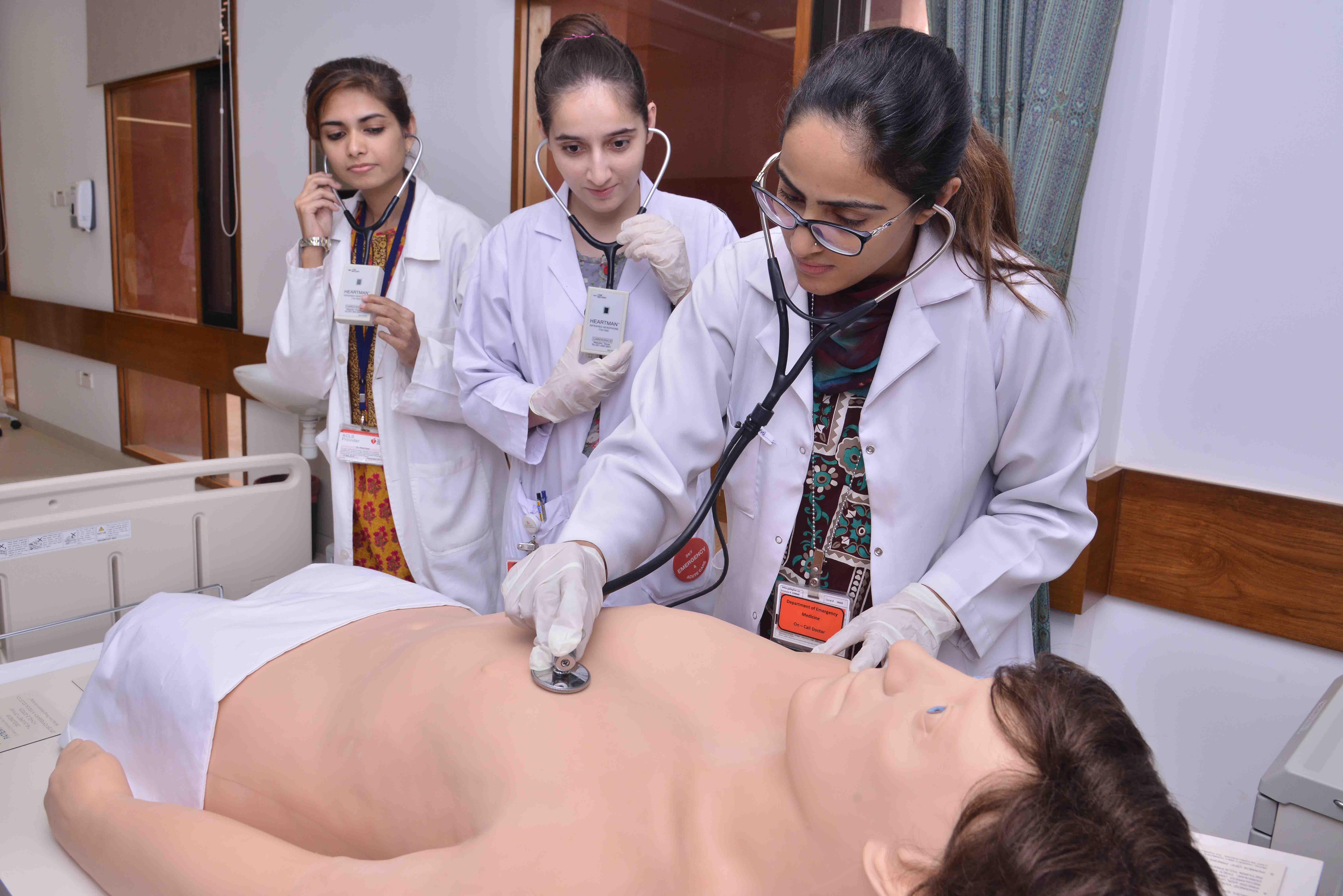
|
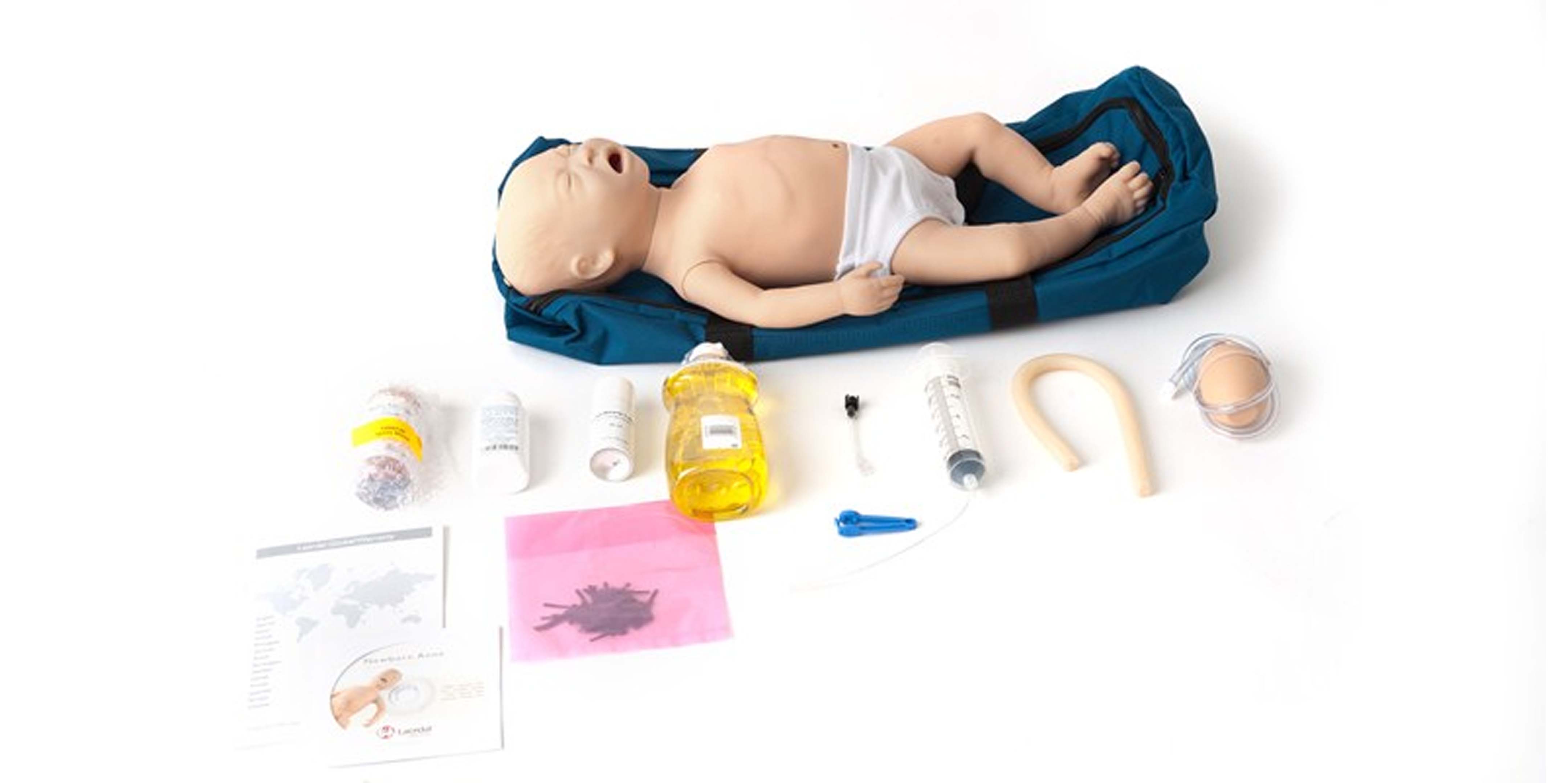
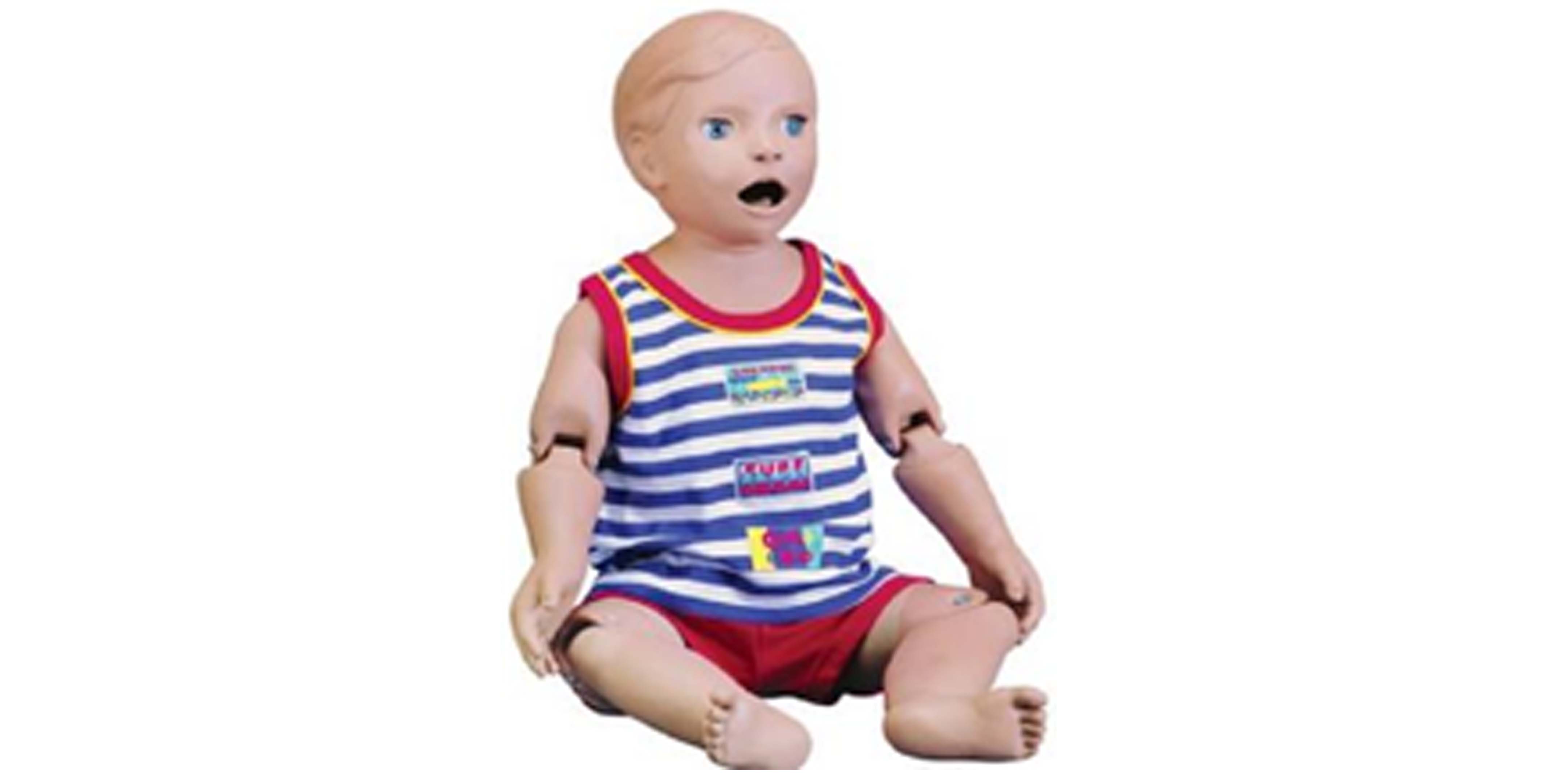
Newborn Anne (Neonatal Mannequin)The mannequin has a realistic airway designed for training in all aspects of newborn airway management, including the use of Positive-Pressure Airway devices, and the placement of ET Tubes and LMA’s. It has realistic chest rise with manual ventilations. The torso includes functionality to relieve a tension pneumothorax via needle decompression. An umbilicus with a manually generated pulse can be assessed, cut and catheterized for IV access. IO access in left and right lower leg, tibial tuberosity and medial malleolus. |
 |
CVC Insertion Simulator IIThe simulator offers efficient training in both landmark and ultrasound-guided central venous catheterization. For both procedures, introductory training is prepareso that trainees can develop their skills in sequence. This is one of a kind simulator that provides simulation of common complications including mislodging.
|  |
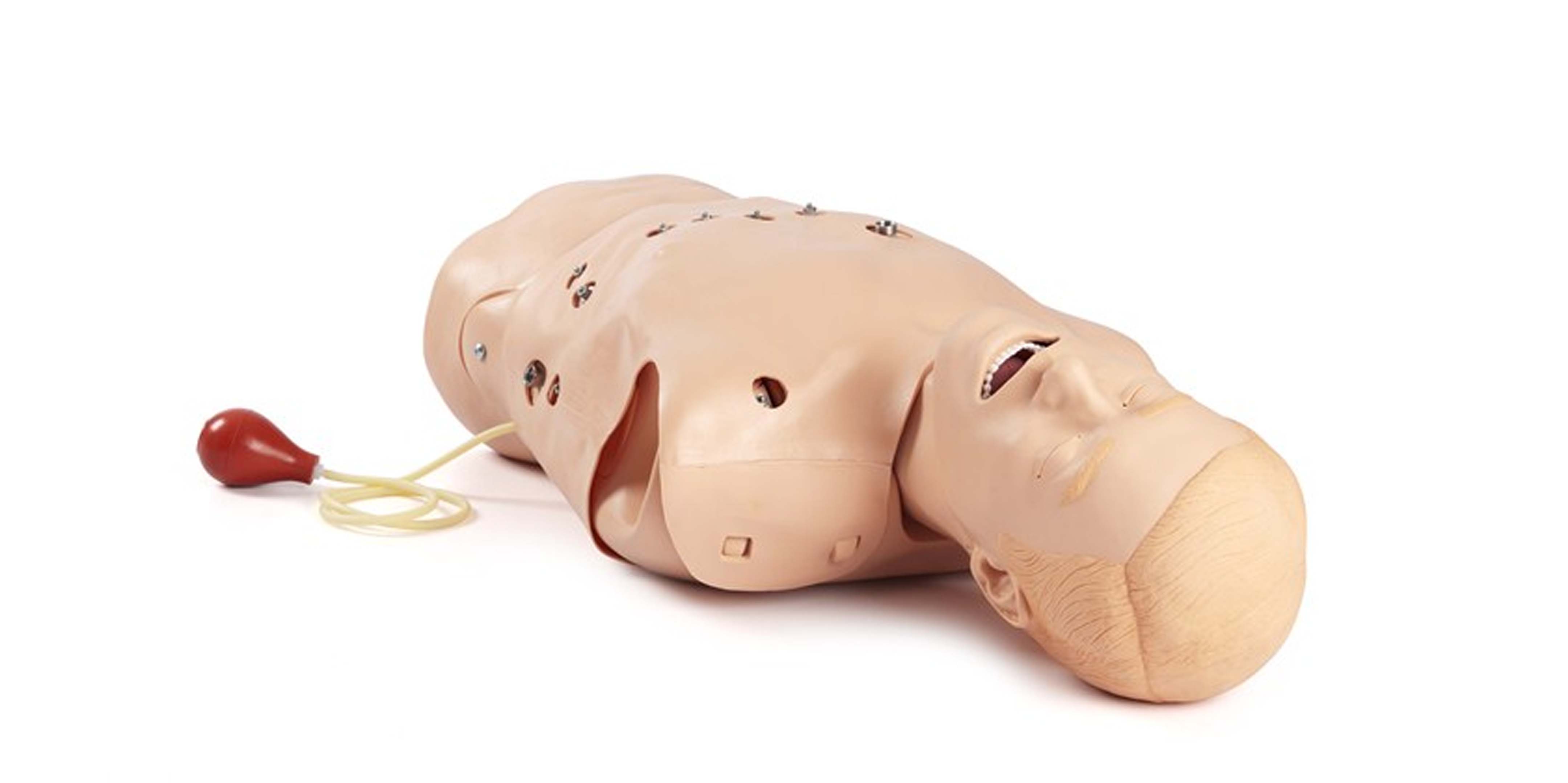
12 Lead Task TrainerAirway management skills:
Updated airway allows for insertion of standard airway devices with the addition of the LMA, Combitube and King LT
Oropharyngeal and nasopharyngeal airway insertion
Manually generated carotid pulse
Stomach auscultation to verify proper positioning
Abdominal Thrust Maneuver may be performed Closed chest compressions
Cardiac-related skills:
Connection sites for four limb leads and VI through V6
Compatible with most 3-, 4- and 12-lead defibrillators/monitors
Pacing with option to respond or fail to capture
Scenarios of up to five (5) ECG choices may be programmed with the ability to step to the next rhythm based on user input time frame or single-button activation
|
 |
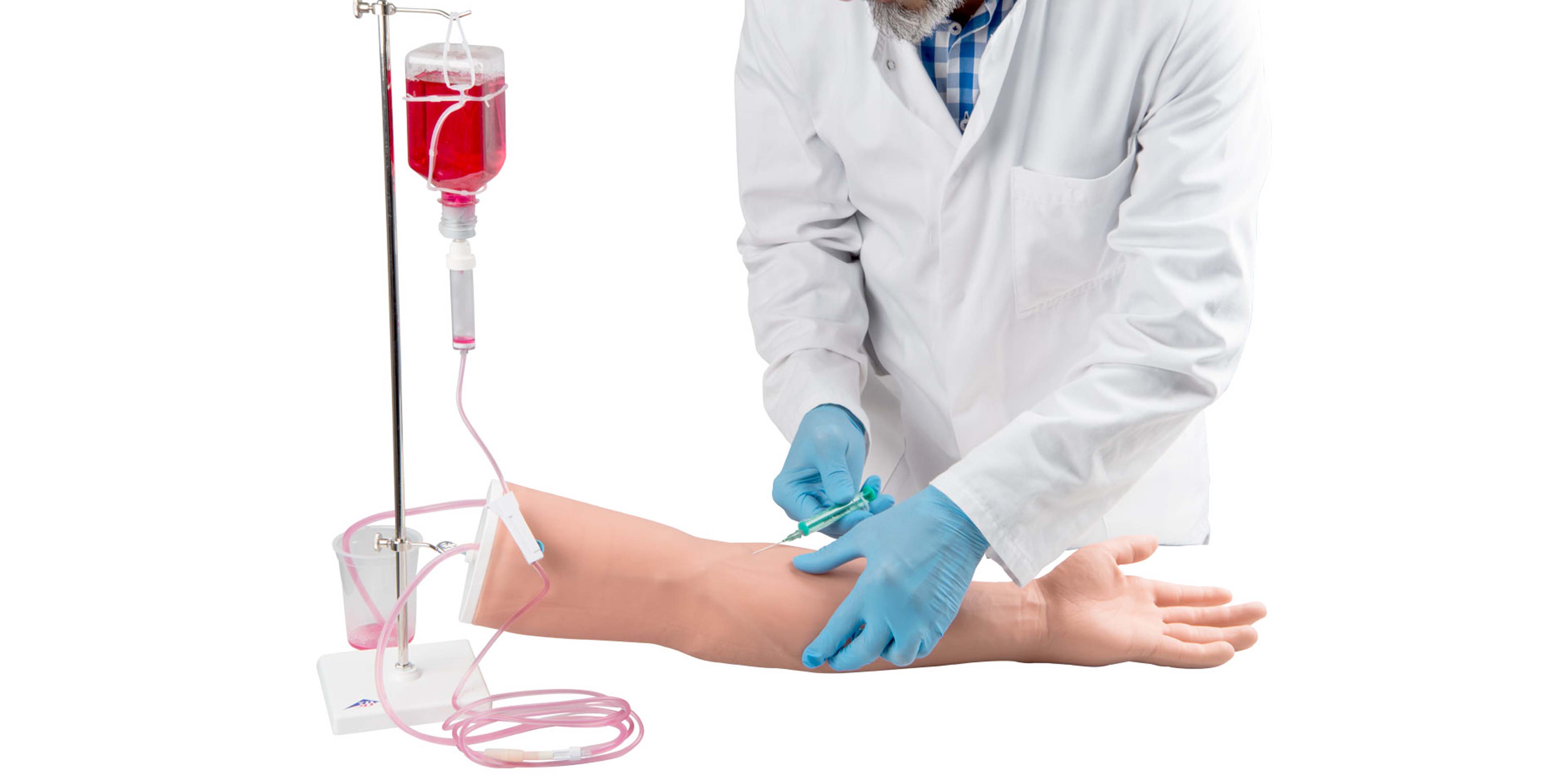
IV Cannulation AdultIt is ideal for practicing Intravenous injections, correct puncture of peripheral veins for blood sampling.
The following veins can be punctured:
Basilic vein, cephalic vein, median cubital vein, dorsal venous rete of hand.
|  |
Basic Life Support Mannequins (Little Family Pack)The Little Family Pack provides quality CPR training. It is designed to reflect the realistic anatomical differences among an adult, a child and an infant.
Our students benefit from the most realistic, age-specific mannequins. The Little Family Pack includes Anne, Junior and Baby Anne. | 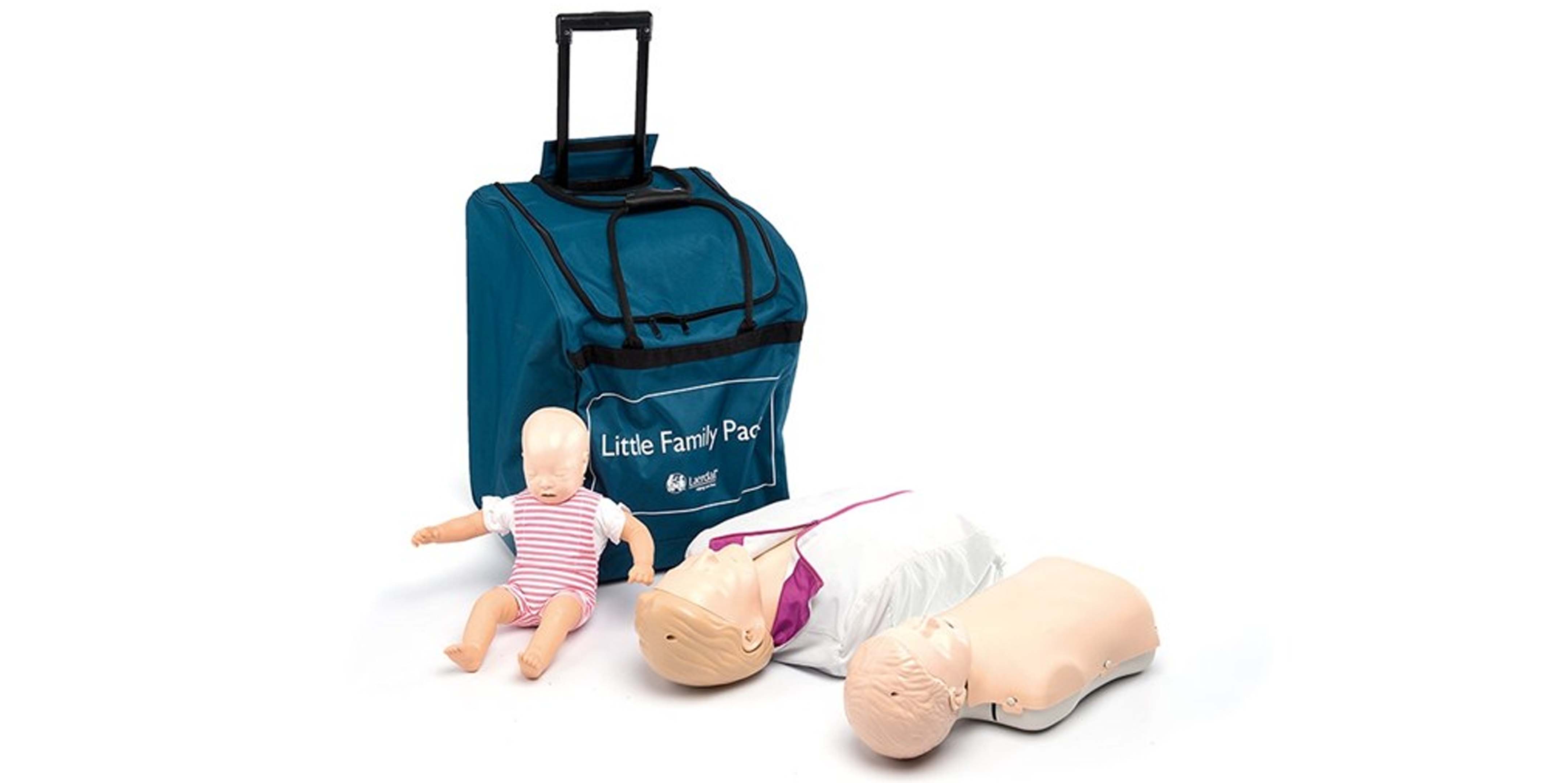 |
NG Adult ModelOne side, covered with a transparent lid, shows the sagittal section including the nose, mouth, pharynx, trachea, esophagus, lungs and stomach. The other side depicts the anatomically correct view of a human torso for teaching the proper method of measuring the NG tube’s length. Torso also features a tracheostomy to demonstrate a variety of care techniques.
|  |
NG PaediatricNG Paediatric has the following features:
Eyes open and close in realistic eyesockets for ophthalmic procedures
Fully articulating head and jaw with teeth and tongue
Jointed elbows and knees
Bathing and bandaging activity
Tracheotomy opening
Male and female catheterization
Enema administration
Oral, nasal, and digital intubation
|  |
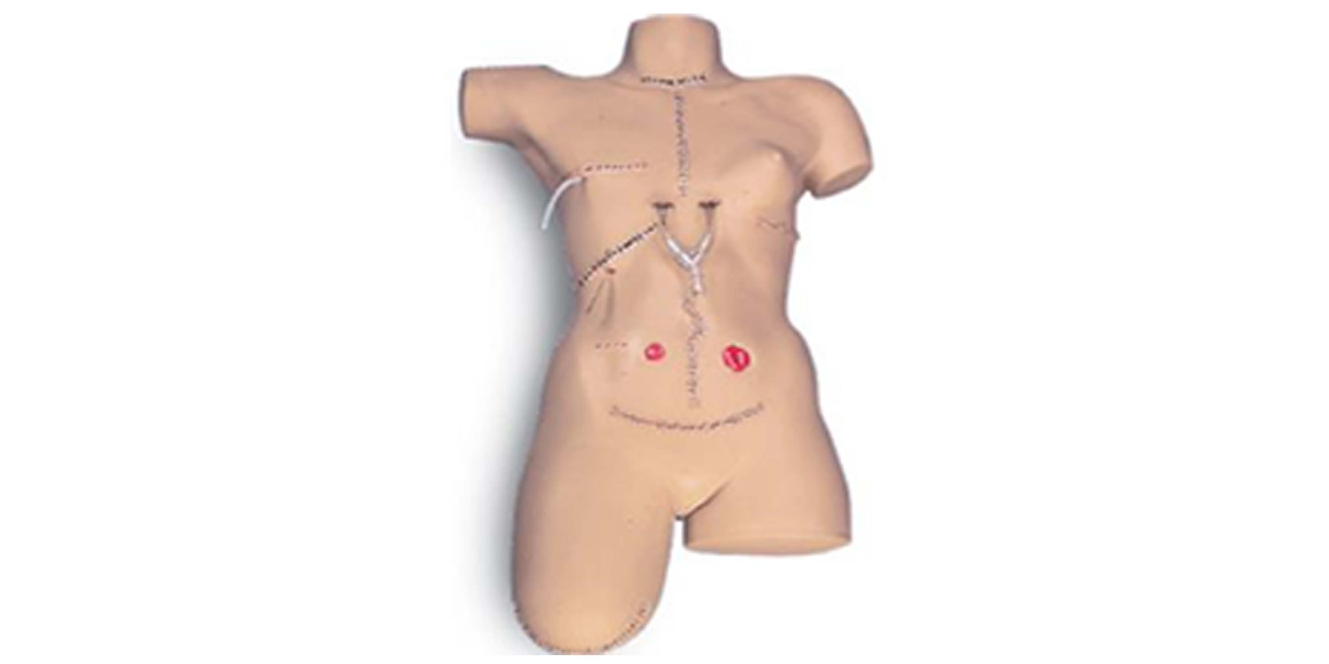
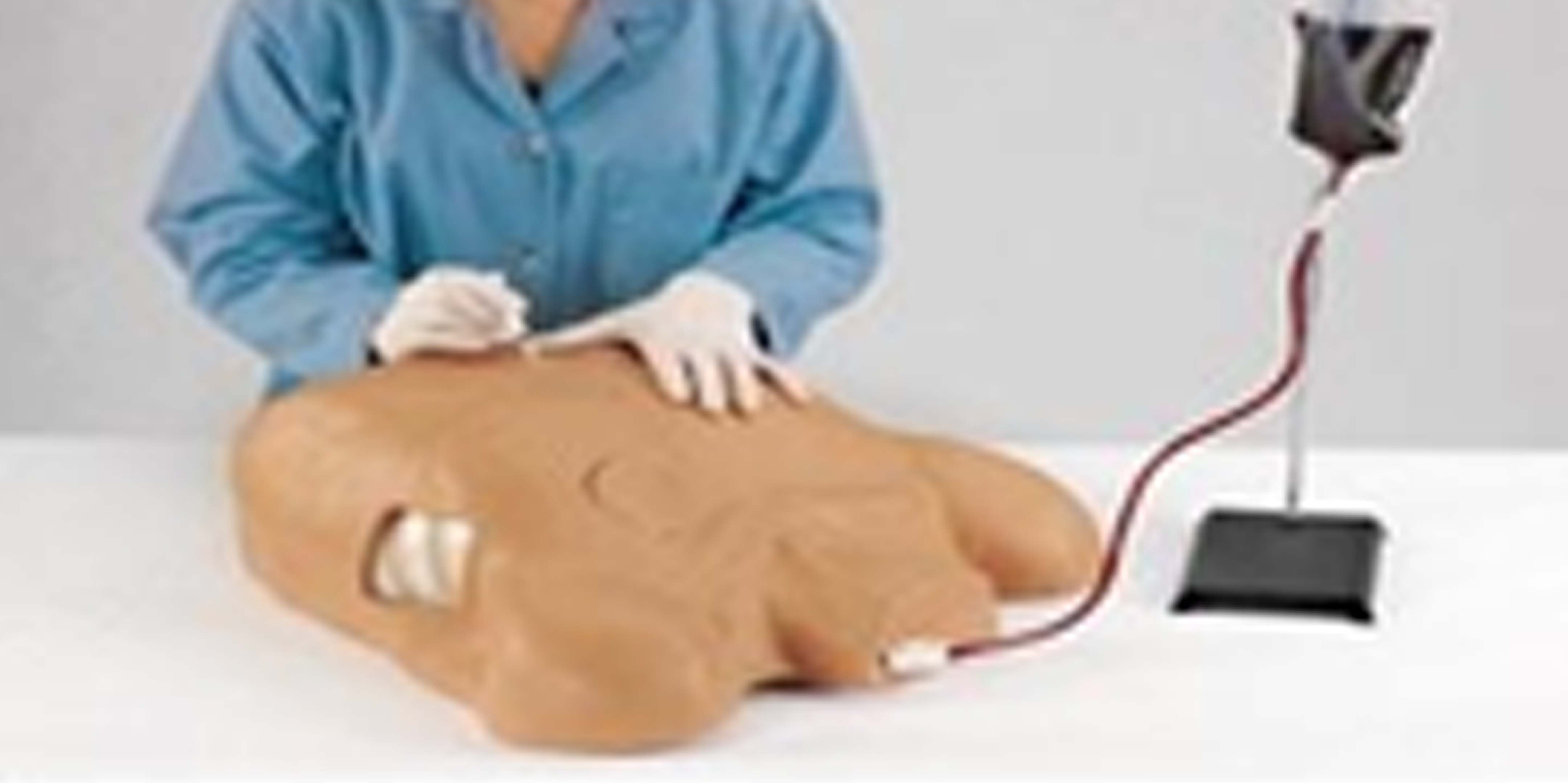
Surgical Bandaging SimulatorSurgical bandaging simulator with 14 surgical wounds including a Mid-Sternal Split with two simulated drains, a Sacral Decubitus Ulcer - Stage 2, and a Leg Amputation Stump. The skin has also been reformulated so it is drier, allowing the bandages to adhere better. Wound closures feature surgical staples for the Mid-Sternal Split, Thoracotomy, Nephrectomy, Laparotomy, Abdominal Hysterectomy, and Amputation Stump. Staples and sutures cannot be removed. It is ideal for teaching wound management skills, dressing changes, and bandaging techniques.
|  |
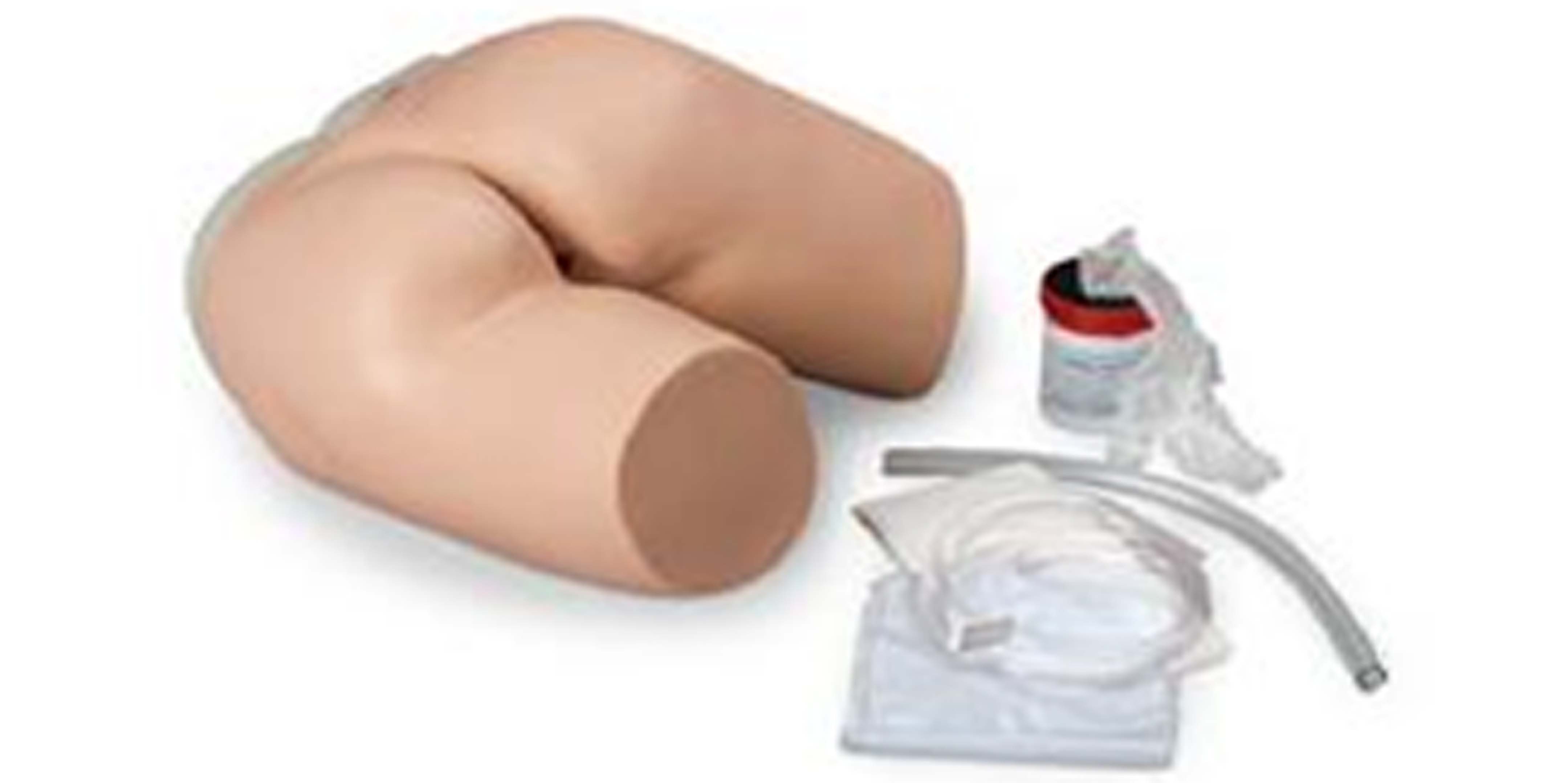
Enema SimulatorThe Enema Administration Simulator is designed to
demonstrate and practice enema procedures. Standard apparatus can be used to introduce fluids.
List of Components
|  |
Epidural and Spinal Injection Trainer It has the following features: Epidural anaesthesia using the loss-of-resistance and the hanging-drop technique
Spinal anaesthesia with realistic resistance of the dura and arachnoid mater with or without a cannula
Fluid-filled spinal canal with realistic outflow rate
An epidural catheter can be inserted into the epidural space
| 
|
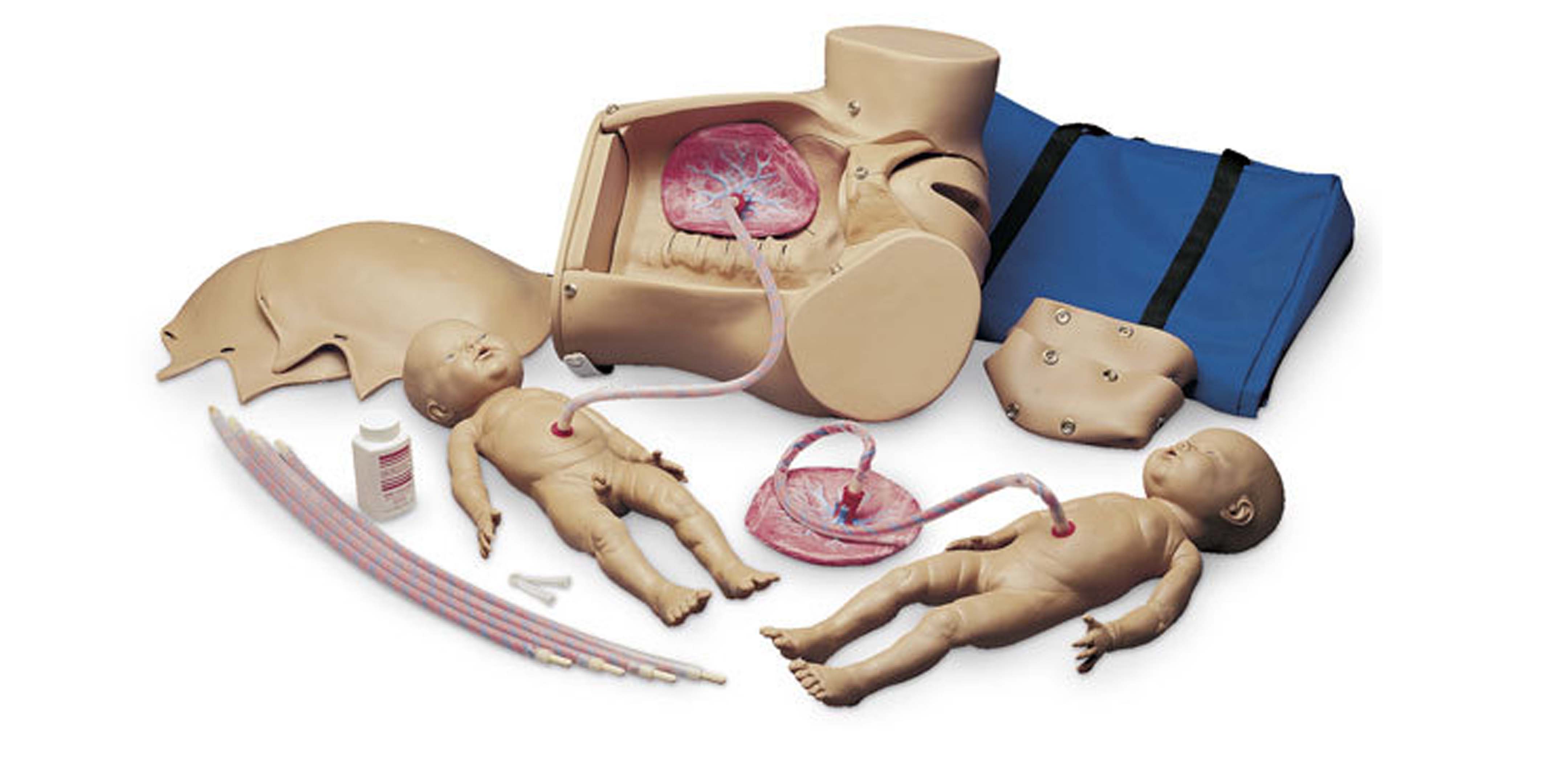
Nita NewBornA unique, anatomically correct representation of a 4-lb., 16" female baby for teaching and practicing the vascular accessing of newborns and infants. Practice accessing, securing, dressing, site care, and maintenance for standard venipuncture, central catheters, PICC lines, and umbilical 5 FR catheters. | 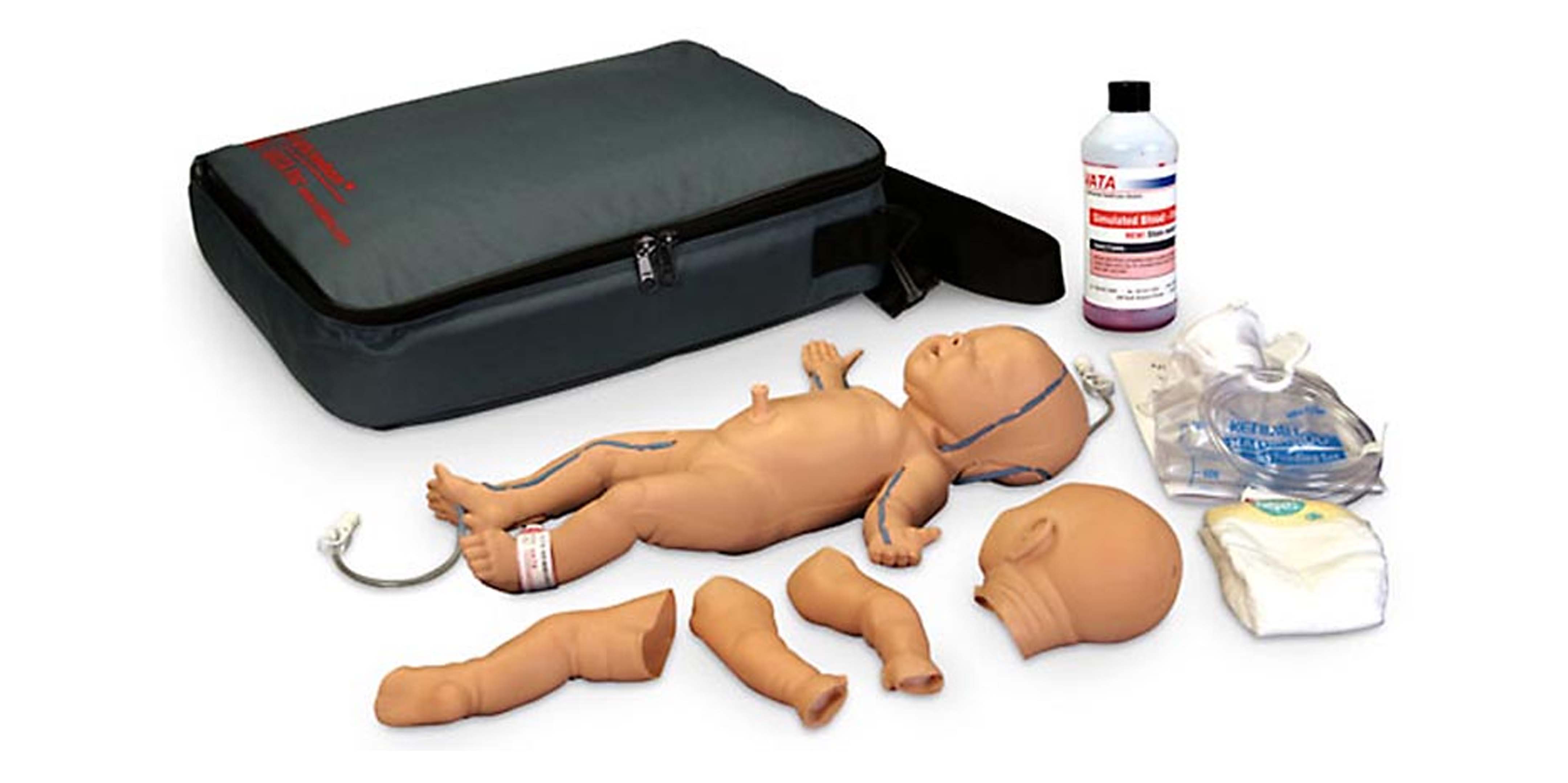 |
MidWifery EpistomyStage 1 (no dilatation, no effacement)
Stage 2 (2 cm dilatation, 50% effacement)
Stage 3 (2 cm dilatation, full effacement)
Stage 4 (5 cm dilatation, full effacement)
Stage 5 (7 cm dilatation, full effacement)
Stage 6 (9 cm dilatation, full effacement)
|  |
MidWiferry Child Birth SimulatorFetal palpation of fontanelles, backbone, knees, and elbows
Vertex presentation
Normal vaginal delivery
Complete, frank and footling breech delivery
C-section delivery
Vertex and breech presentations in multiple delivery
Demonstration of placenta previa: total, partial, and marginal Prolapse of umbilical cord
Ritgen's maneuver
3 soft vulva inserts for episiotomy exercises
Normal delivery of umbilical cord and placenta
Suction of baby's nose and mouth
|
 |
Pelvis Trainer with all accessoriesAnatomically accurate perineum and pelvic floor muscles, with bony pelvis modeled from CT scan data. The abdominal skin can be removed to display anatomical structures and fetal positioning. Representative full term baby with palpable fontanel and suture lines, palpable clavicles and scapulae, and detachable placenta with cord. Mother allows for multiple delivery positions and techniques: |
 |
Eye Examination Simulator
It is an innovative trainer for fundus examination, designed to allow examination of eyegrounds with the physician's own ophthalmoscope.
Various cases can be set up for trainees using combinations of choice of slides, depth and pupil diameter.
| 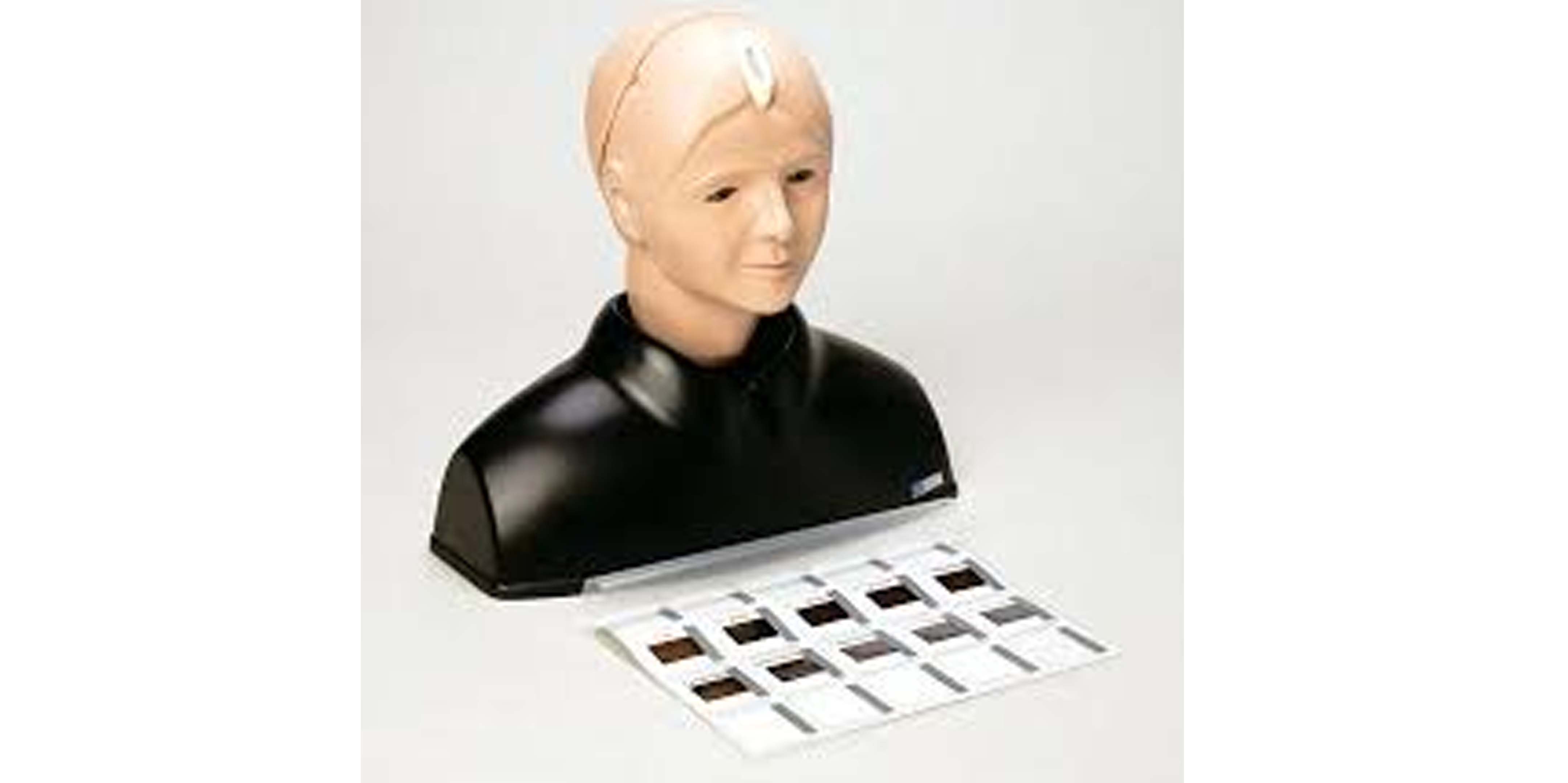 |
Ear Examination SimulatorA life-size model of the ear including external acoustic meatus and tympanic membrane, for training in examination of the ear with an otoscope and foreign body removal. It offers 2 modes of use: instructor mode, which is is particularly useful for OSCE or skills examination, and self-learning mode.
| 
|
Breast Cancer Training ModelIt offers unparalleled realism for teaching both clinical and self-breast examination. Unlike other simulators, tissue density actually varies within the simulated breast, just as it does in a live patient. Tumors of varying sizes (1-4 cm diameter), shapes (round, oval, irregular/stellate), and densities can be inserted by the instructor for an expanded combination of training scenarios. | 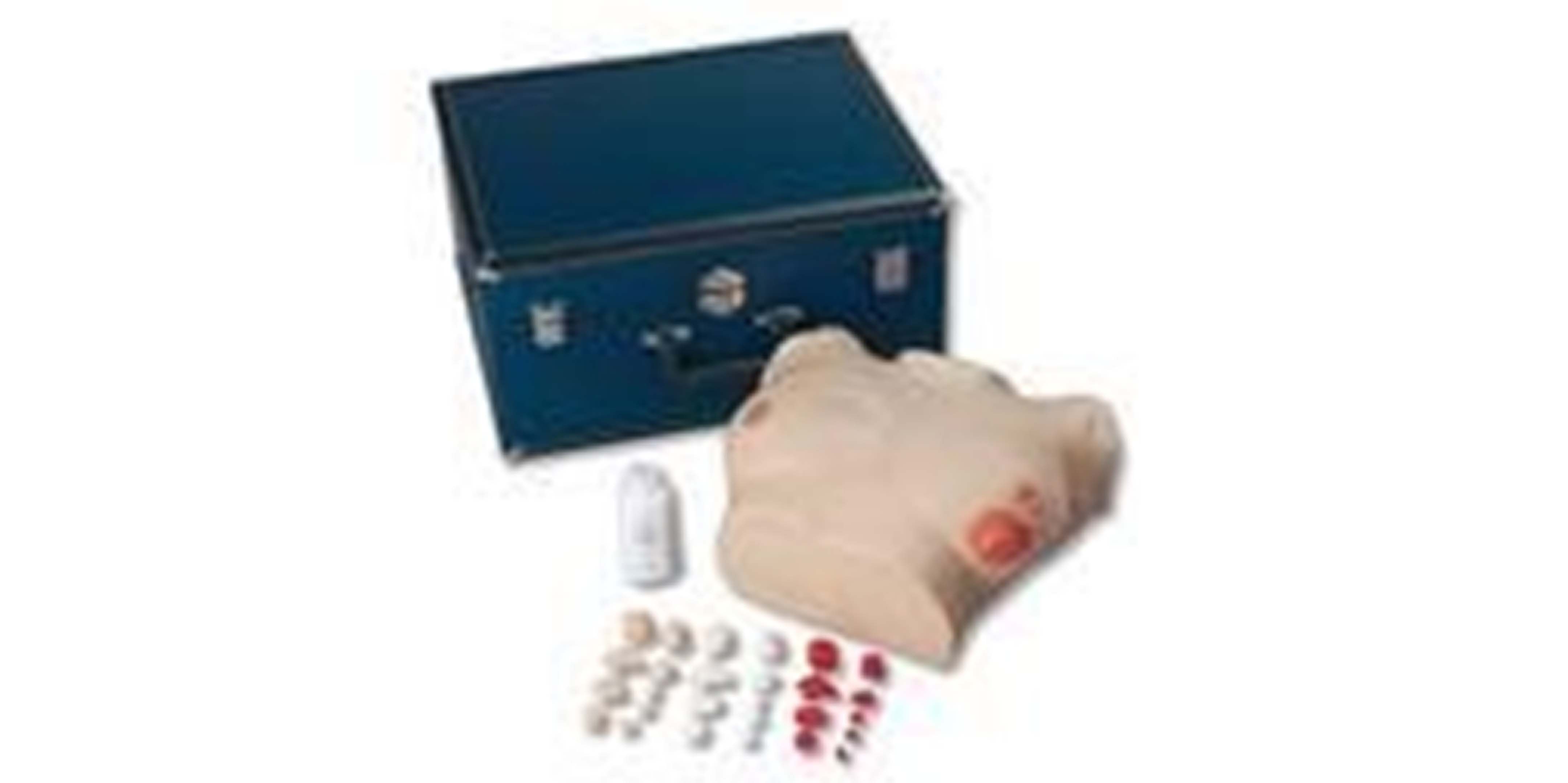
|
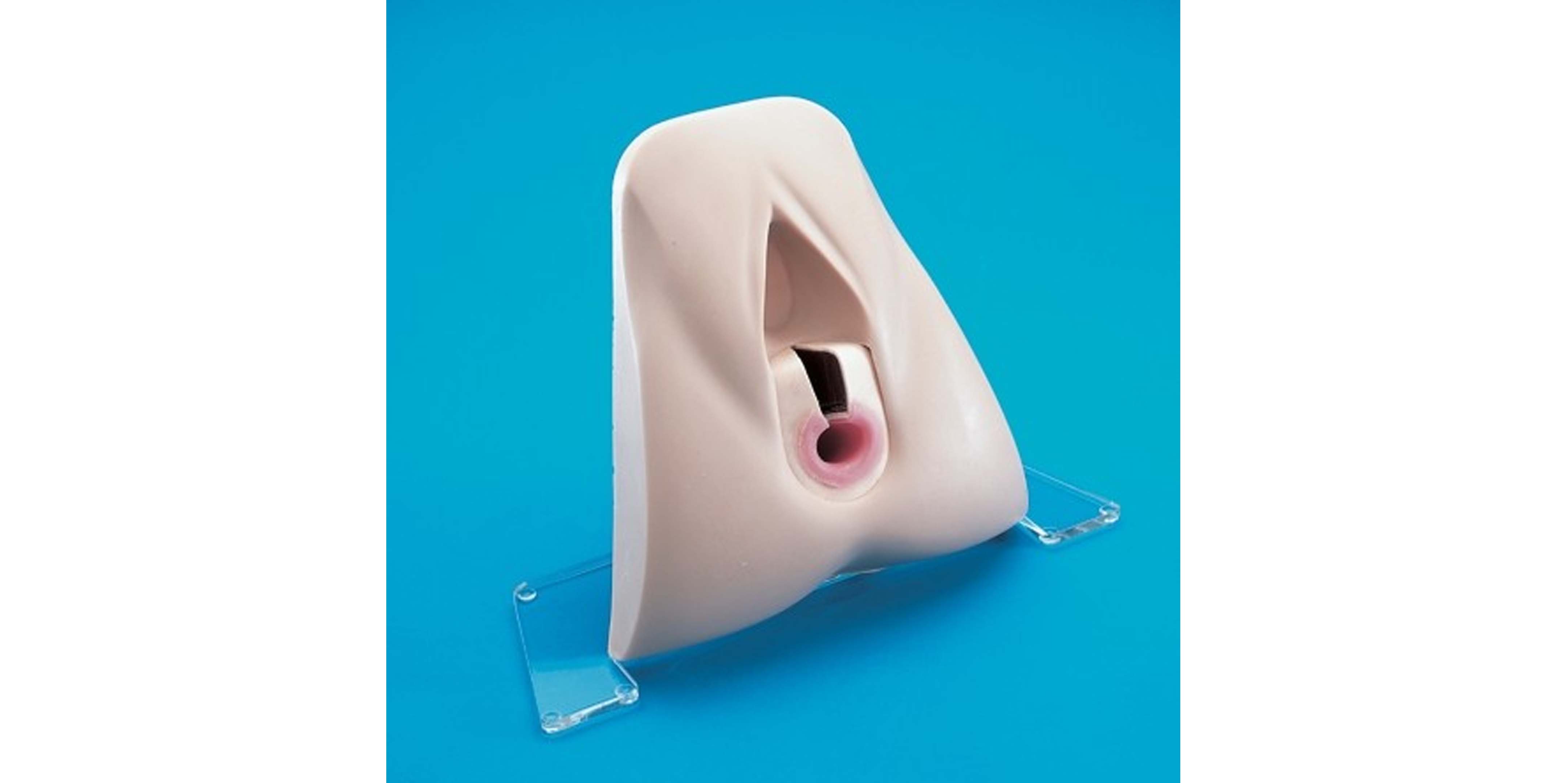
IUD TrainerThis anatomically accurate model represents a section of the uterus, ovaries and fimbrae. The uterus is covered by a clear plastic window to allows easy visualization of insertion and placement of IUD. |

|
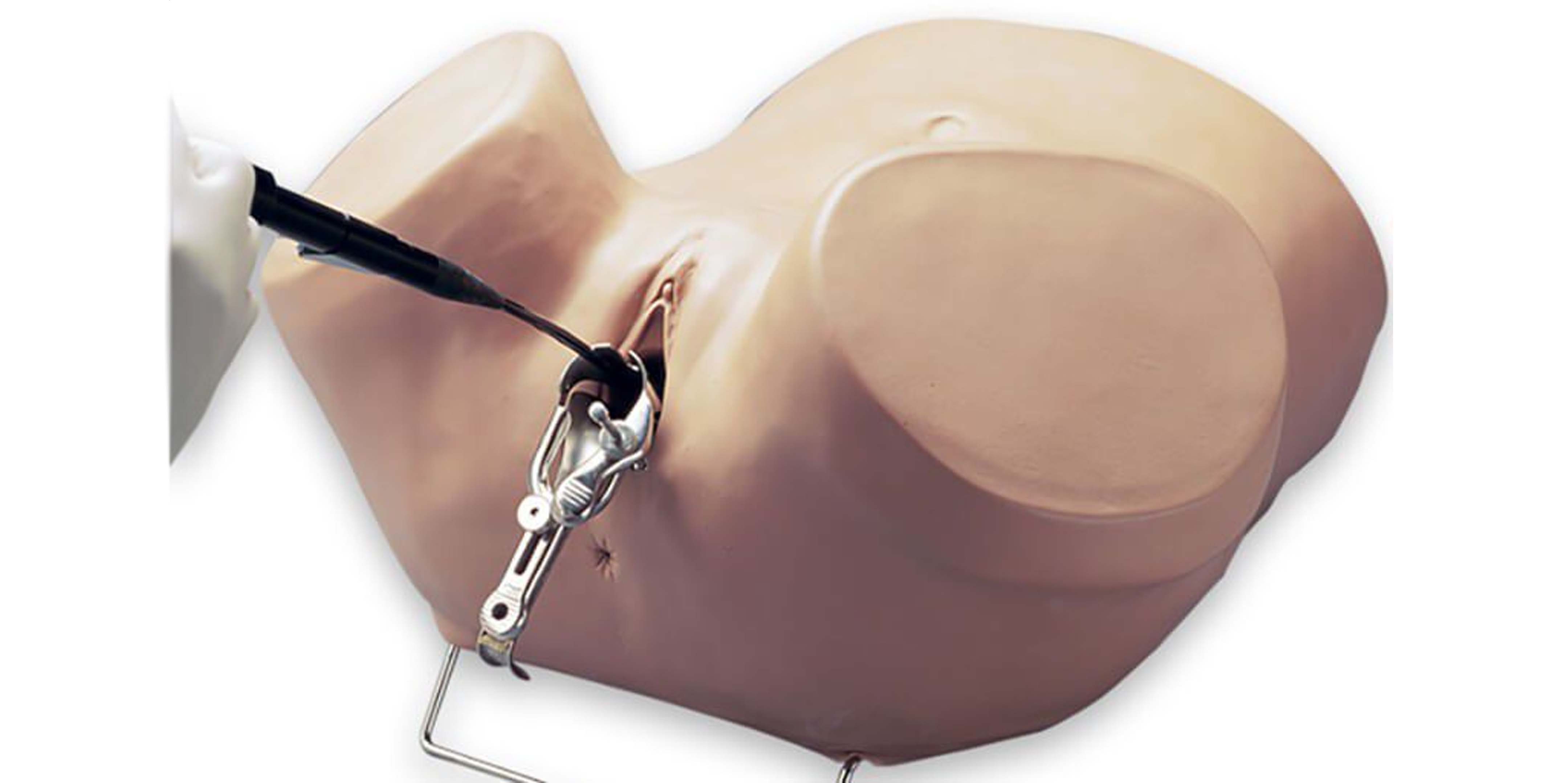
Sultan Anal Sphincter TrainerFor practicing the techniques associated with the repair of the internal and external sphincter.
| 
|
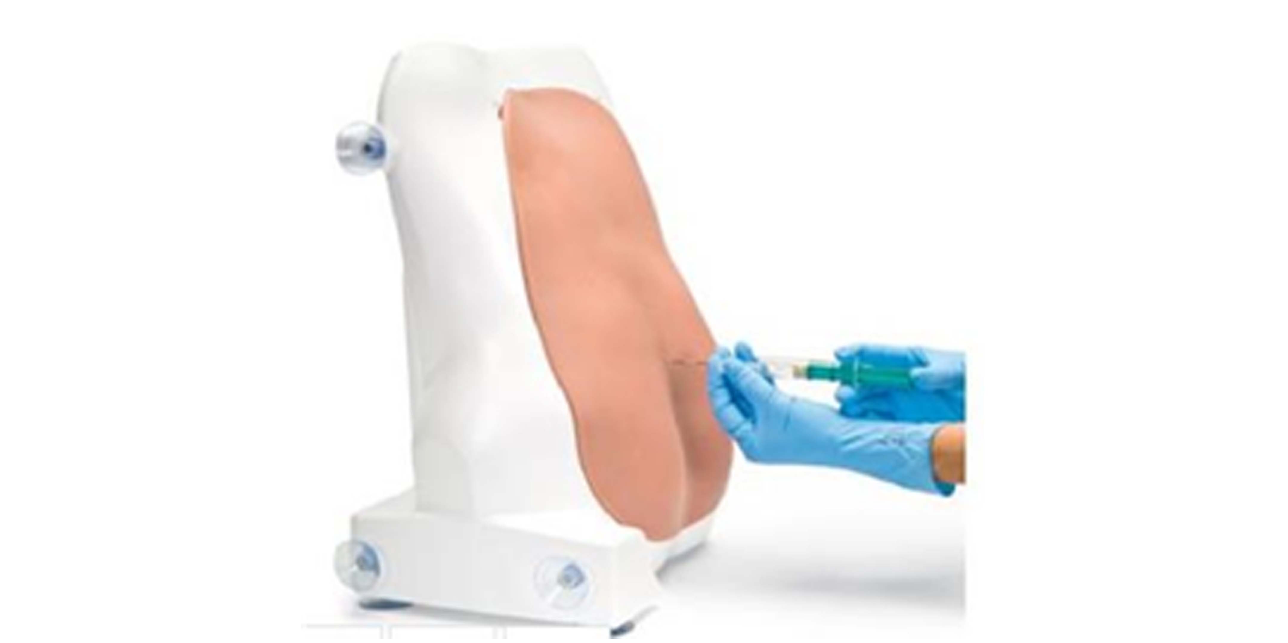
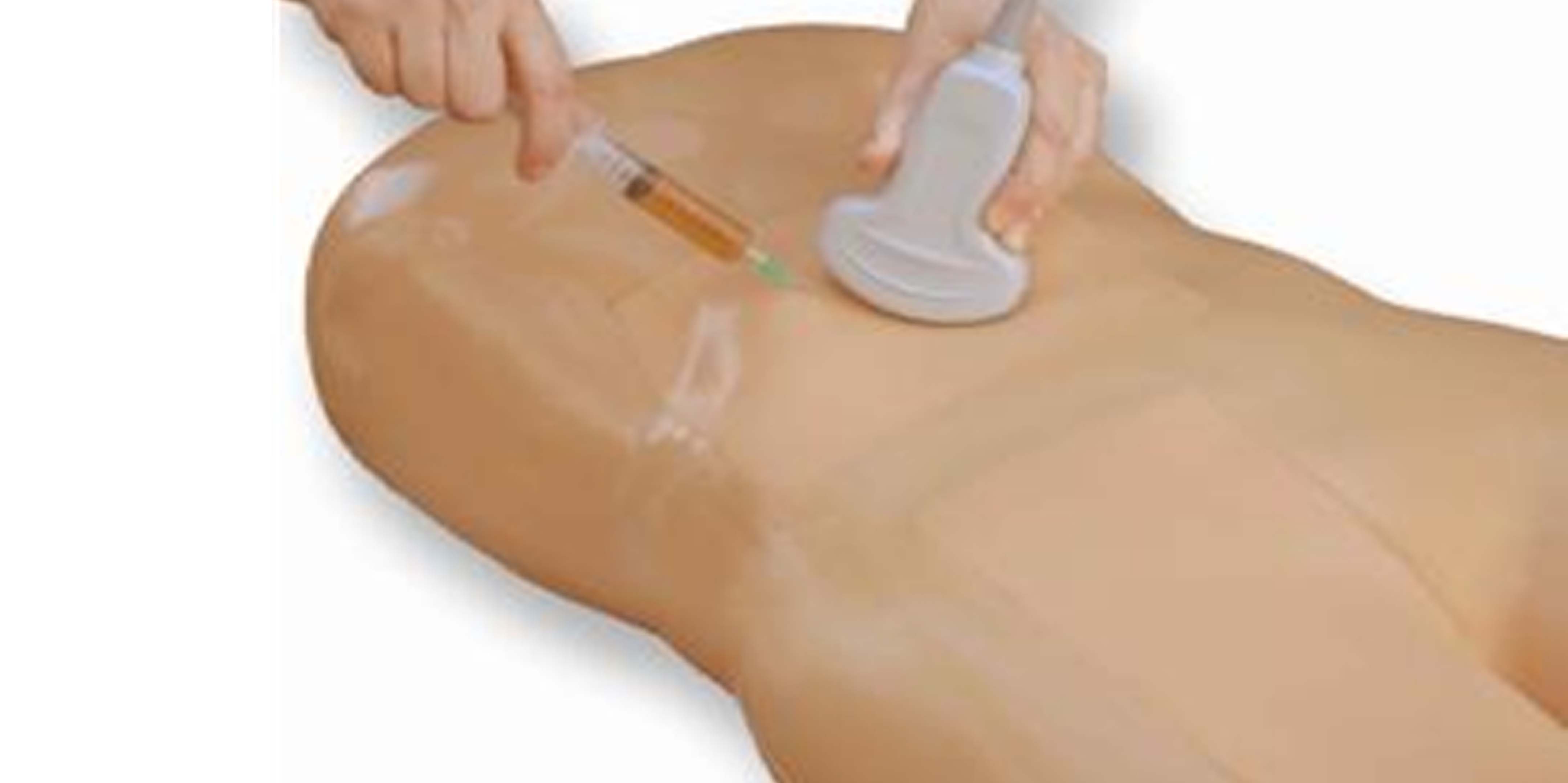
Training Arm for Implantable Hormonal ContraceptivesRepresentation of an upper left arm with soft foam insert to train insertion and removal of implantable hormonal contraceptives such as Levonorgestrel.
|  |
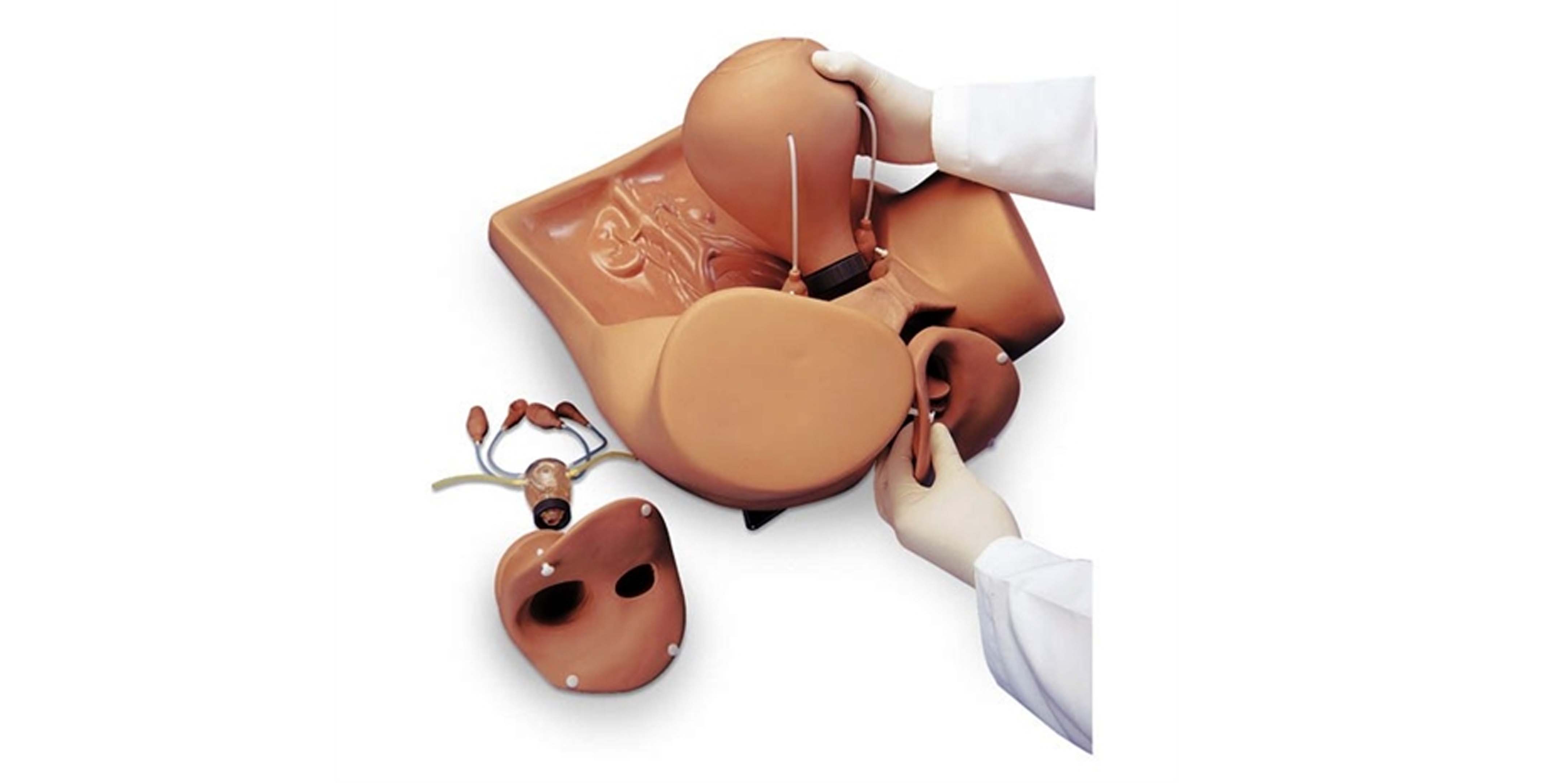
Hysteroscopy Diagnostic TrainerA full size female adult lower torso with a foam-backed soft outer skin simulates the feel of the anterior pelvic wall.
Features includes:
- Full size female adult lower torso with removable soft outer skin
- Seven uteri including normal “control” uterus with healthy internal and external appearance
- Internal anatomic artwork of normal and abnormal uteri
- Internal representation of endometrial polyposis, fibroids, early and advanced carcinoma of the endometrium, torsion of sloughing fibroid, and carcinoma of the fundus
| 
|
ZOE Gynaecologic SimulatorZOE combines the ability to demonstrate multiple gynaecologic procedures as well as practice laparoscopic examination and minilaparotomy. The features includes: Full size adult female lower torso with relevant internal anatomic landmarks Palpation of normal and pregnant uteri Vaginal examination, including
insertion of speculum IUD insertion and removal Laparoscopic visualization and occlusion of fallopian tubes
|
|
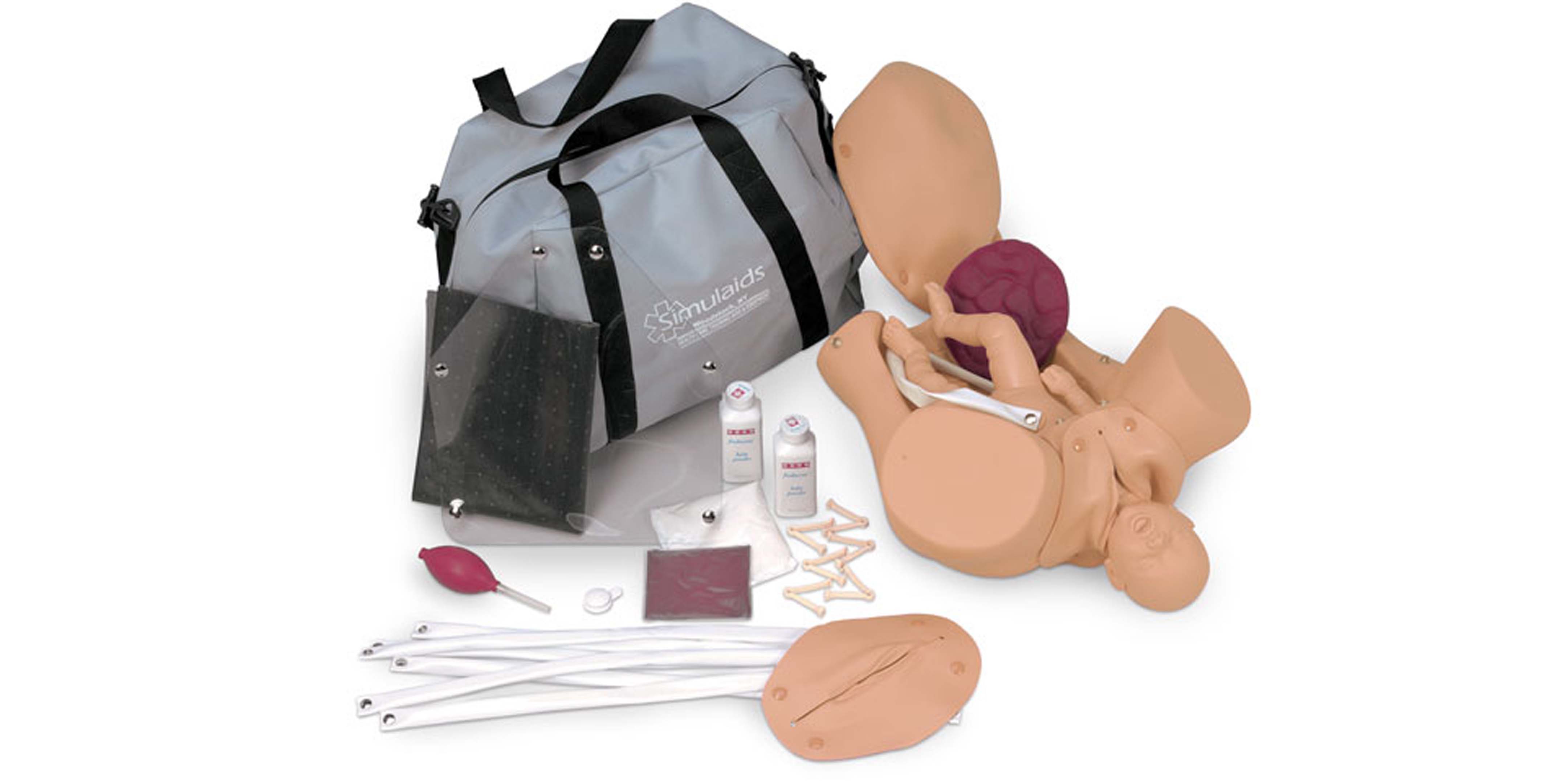
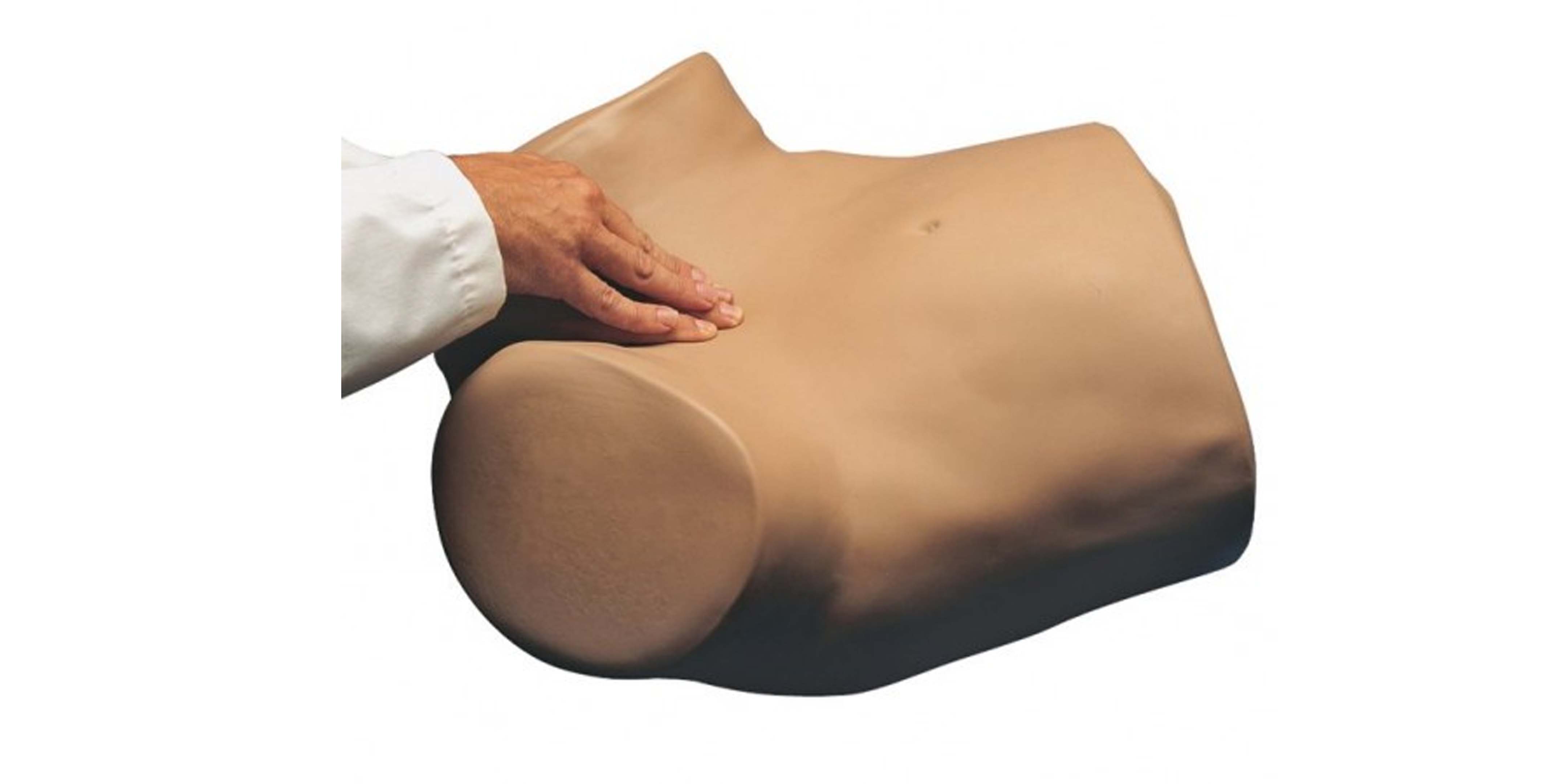
Obstetric ExaminationAnatomically correct pelvic model with full-term newborn and placenta to
give realistic practice in multiple techniques and to learn the
procedure for emergency childbirth. Disposable umbilical cords with clamps, easily replaced extra vulva,
powder to make simulated blood, a modular pregnant belly overlay with
permanently installed fetus for training practice of Leopold’s maneuver
to determine the fetus’ lie by palpating its skull and kneecaps, and a
clear abdominal overlay to see positioning are all included.
| 
|
Gynaecologic Simulator
The full size gynecological simulator of a
female adult lower body gives students and educators a graphic
experience in vaginal speculum examination, bi-manual pelvic
examination, IUD insertion techniques, diaphragm sizing and fitting,
uterine sounding, and viewing of normal and abnormal cervices.
|  |
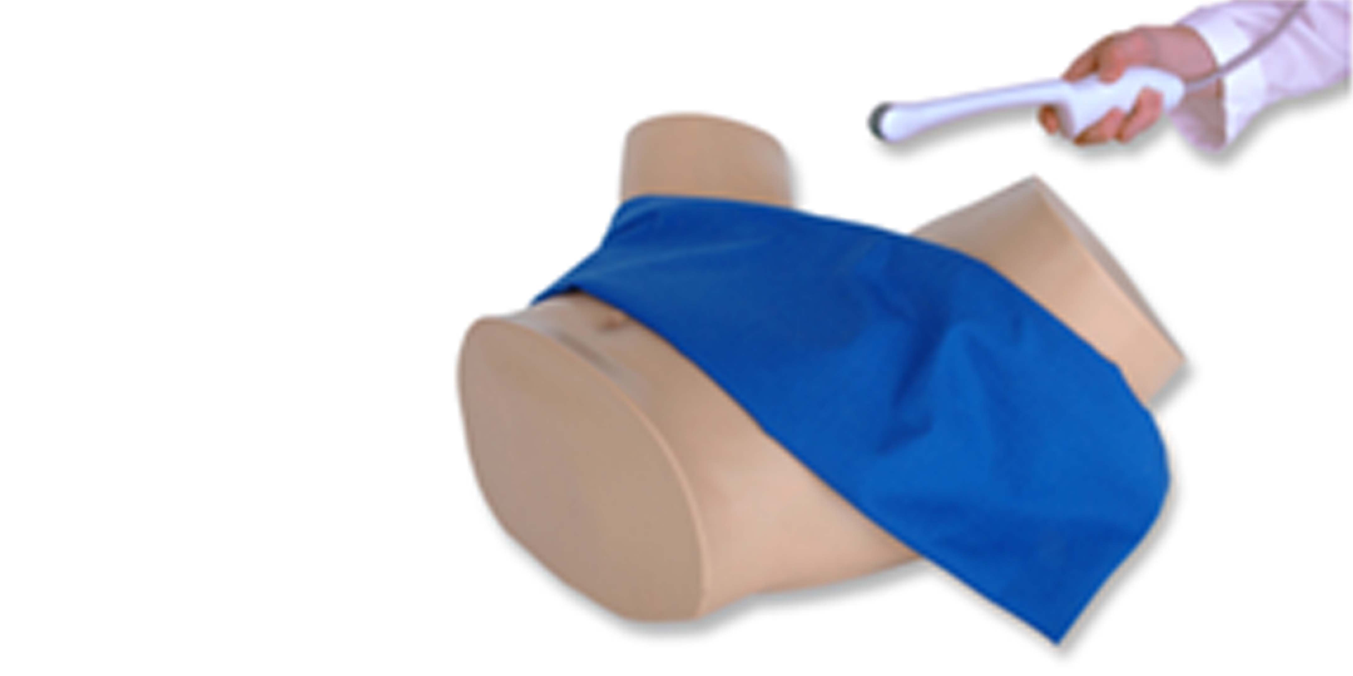
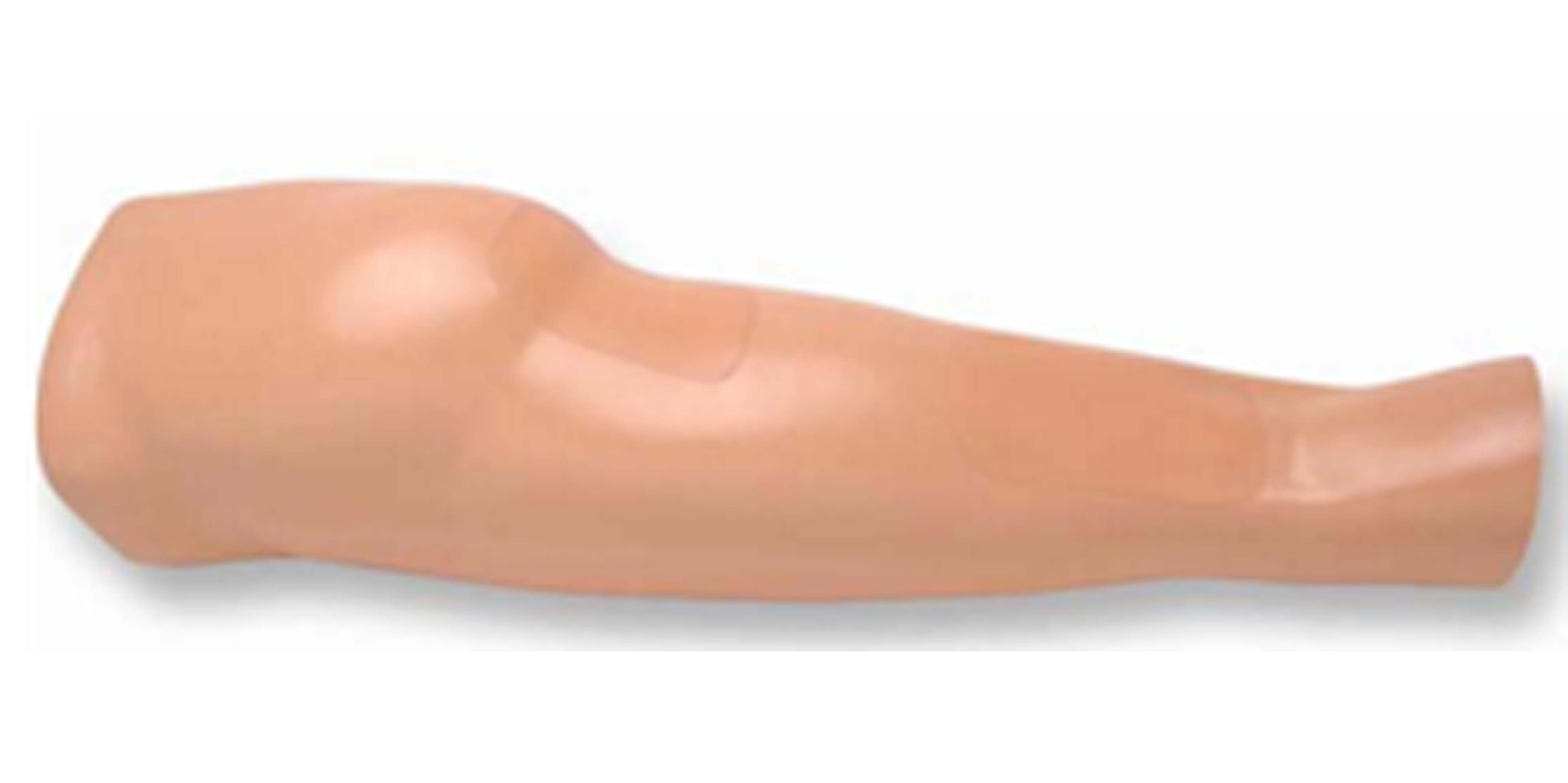
Arterial Arm Stick Kit
Lifelike adult male arm reproduction with infusible arteries for training the proper arterial puncture procedure for blood gas
analysis. Arm has injectable deltoid pad and fits adult male manikins. | 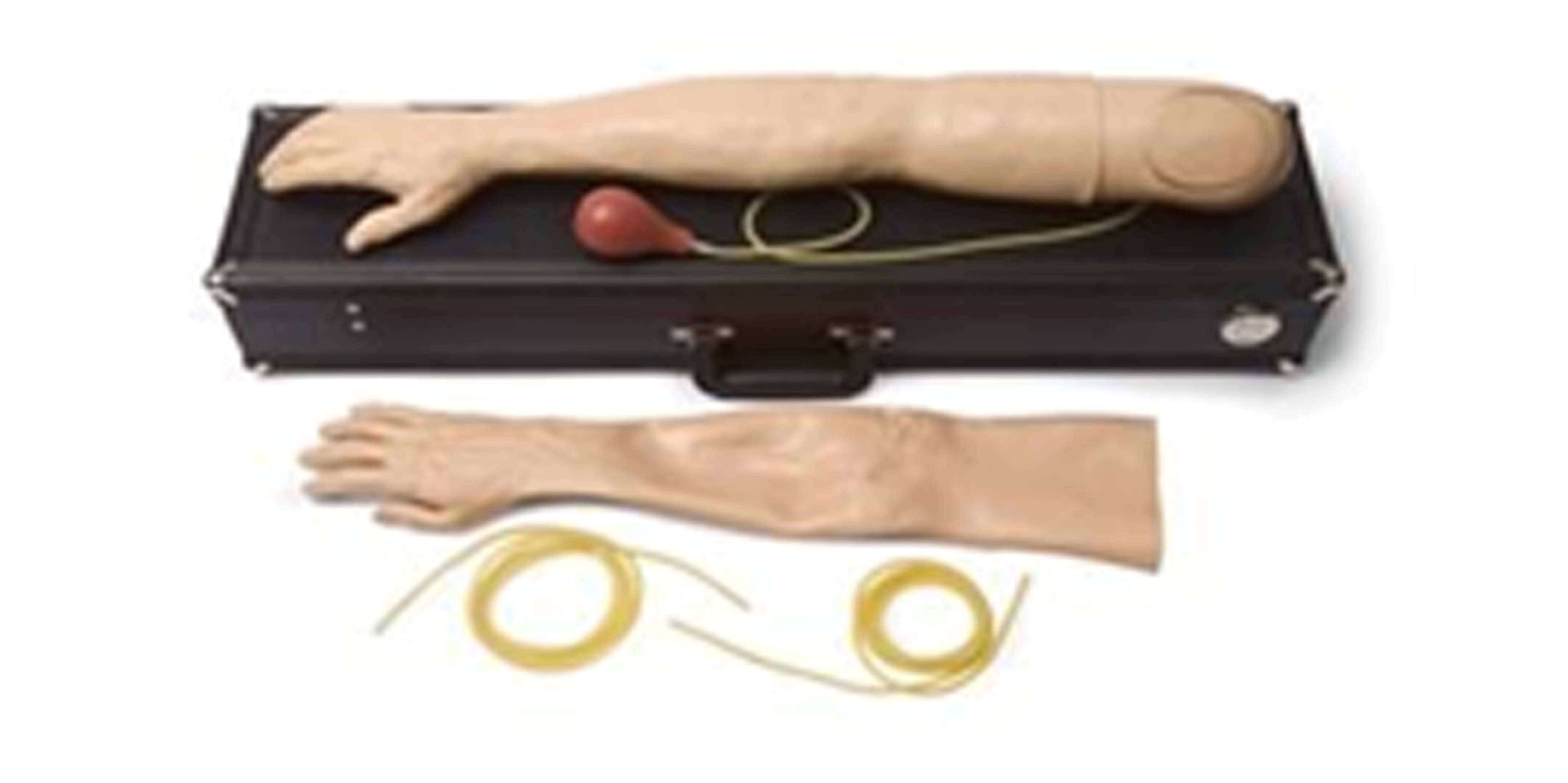
|
Neonate for Umbilical artery CannulationUmbilical Catheterization Site | 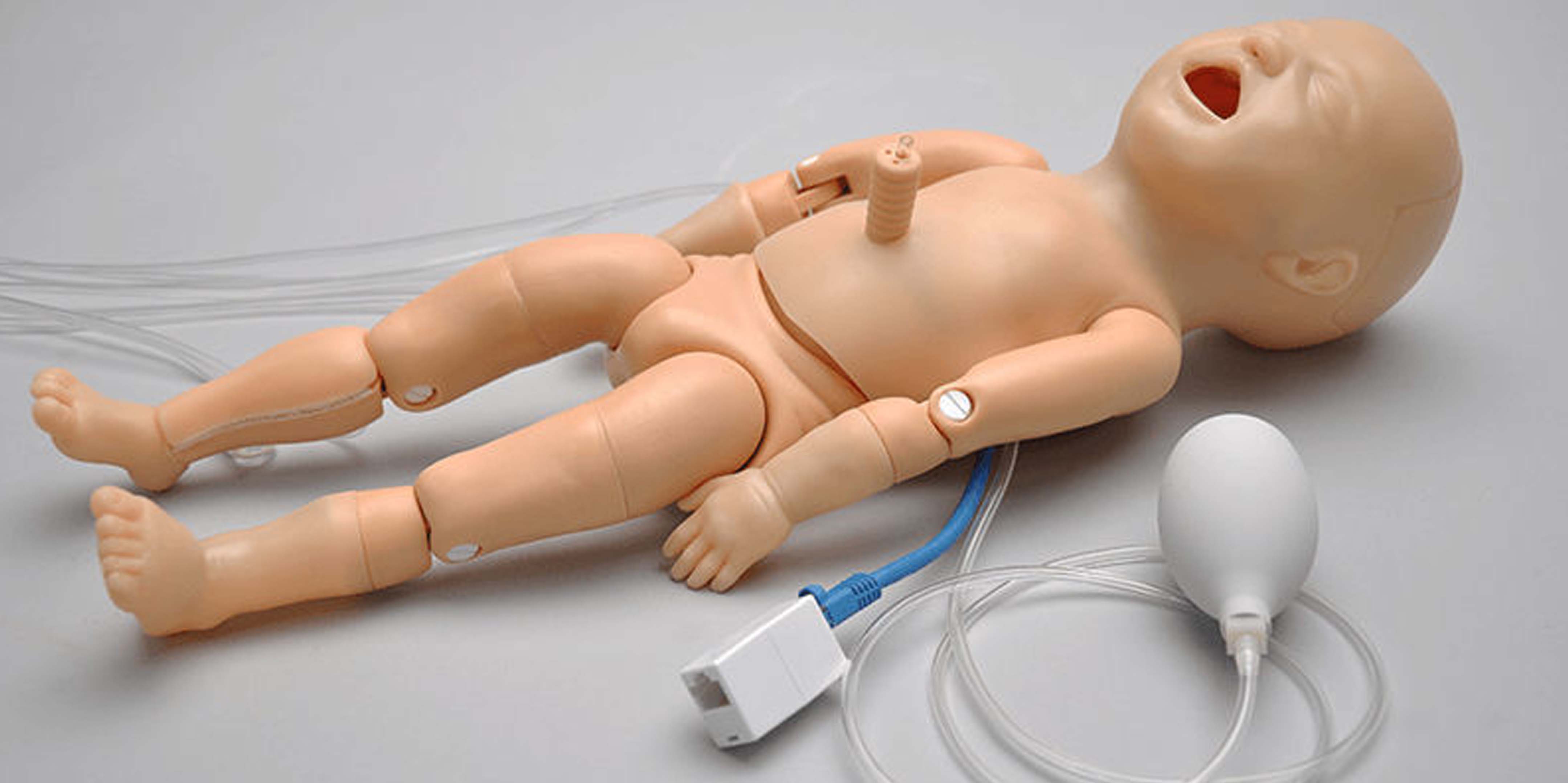
|
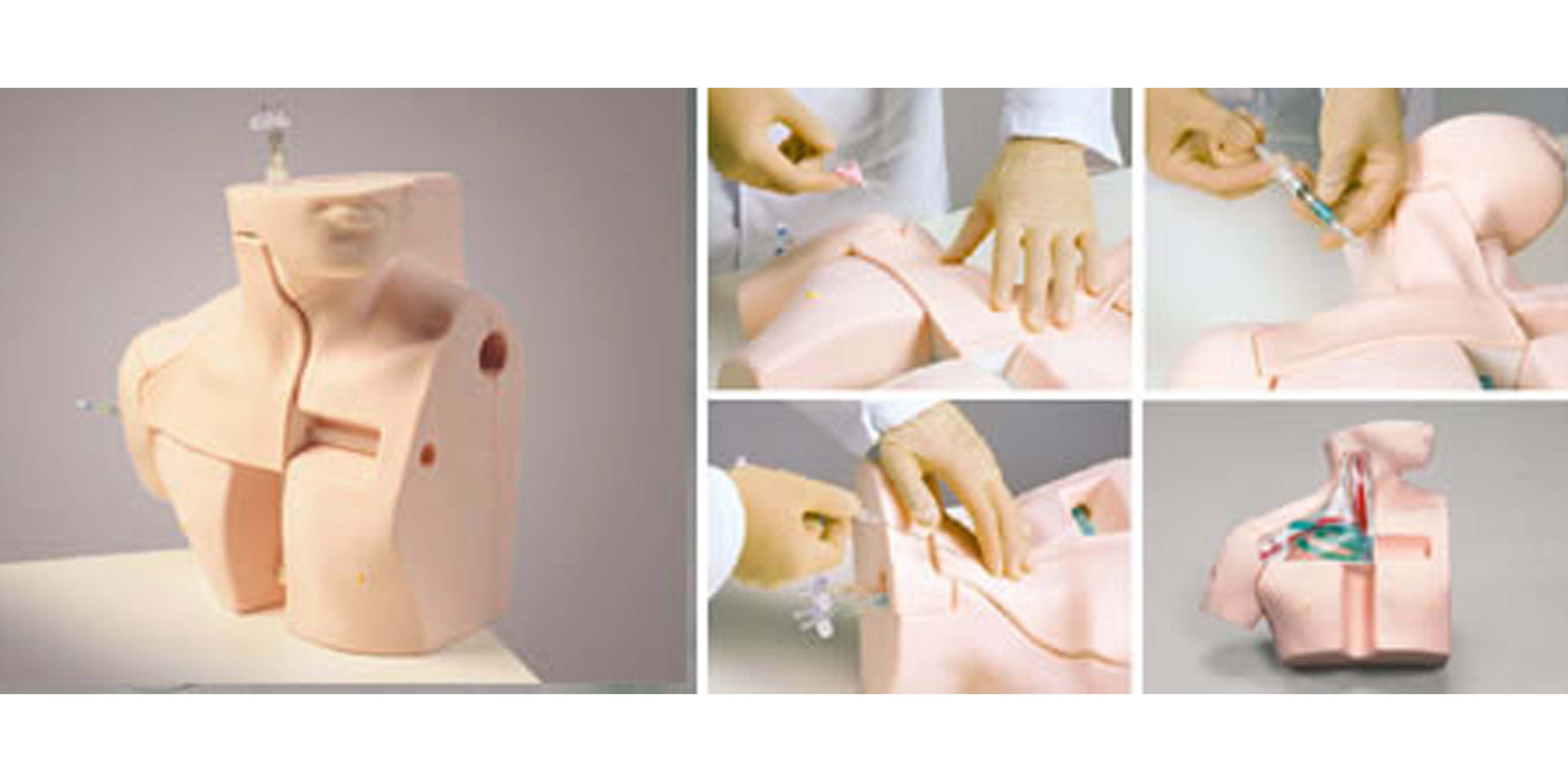
PeriCardio Centesis SimulatorIt is designed specifically to teach the skills needed to perform this difficult procedure correctly, as well as ongoing chest tube maintenance and the management of pre-hospital chest trauma. It has a pressurized tension pneumothorax site and a site for the surgical placement of a functional chest tube. Fluid color, volume, and viscosity can be controlled by the instructor. Pericardiocentesis may be performed in the left subxiphoid space and in the left fifth intercoastal space.
| 
|
Cardiac Auscultation Simulator
Heart & Breath Sounds Simulator
| 
|
Paracentesis Ultrasound Training ModelRealistic and ultra-durable ultrasound guided paracentesis model for training clinicians in the psychomotor skills associated with ultrasound guided paracentesis procedures.
Ultra-durable self healing tissue is extremely realistic in ultrasound imaging characteristics and feels like real human tissue.
Anatomically correct anatomy of the mid abdomen including the right lobe of the liver, small bowel, transverse colon, and peritoneal fluid collections.
|
|
Transvaginal UltrsoundGeneral Pathology Transvaginal Ultrasound Training model
|  |
Nerve Block Upper Limb ManikinRegional Anesthesia and Ultrasound Central Line Training Model
Blue Phantom’s upper torso ultrasound guided regional anesthesia and central venous access training model is excellent to train users to develop and practice the skills necessary to gain proficiency in using ultrasound for peripheral nerve block regional anesthesia procedures of the brachialplexusas well as guide catheter insertions in the internal jugular vein (IJ), subclavian vein, and axillary vein.
| 
|
Nerve Block Lower Limb ManikinSciatic Nerve Leg Regional Anesthesia Ultrasound
Training Model
Blue Phantom’s femoral lower torso ultrasound guided regional anesthesia and central venous access training model is excellent to train users to develop and practice the skills necessary to gain proficiency in using ultrasound for regional anesthesia procedures of the femoral nerve as well as guide catheter insertions in the femoral artery and vein. The DVT option allows users to learn to identify deep vein thrombosis of the deep veins of the legs.
| 
|
Ultrasound Thoracentesis
The Ultrasound Thoracentesis Model simulates a partial torso with anatomical landmarks including the scapula, ribs, diaphragm, pleural cavity, and lung. A positive fluid flow then offers users feedback when pleural effusions are accurately accessed. The trainer is designed to be filled with fluid and is self-sealing for multiple procedures.
| 
|
Arrhythemia SimulatorThe following 6 different arrhythmias are available for pacer training: Sinus
Brady, Junctional Brady, 2nd degree type I A-V block, 2nd degree type
II A-V block, 2nd degree type II A-V block with PVCs and 3rd degree A-V
block. Cardioversion can be simulated by defibrillating the manikin with a manual, semi-automatic, or automatic defibrillator.
The
following 11 arrhythmias are available for defibrillator training: V.
Fib, V. Tach (fast), V. Tach (slow), V. Tach (polymorphic), A. Fib, A.
Flutter, SVT, Sinus Tach, Sinus rhythm with PVCs, Asystole and NSR. An
LED illuminates for: pacer pulse detection, defibrillator discharge
detection, adult mode, pediatric mode and low battery. Battery saver
feature powers-off simulator automatically when not in use.
|
|
Arrhythemia SimulatorCPR
Palpable and visual landmarks
Fully articulated head, neck and jaw
Airway Management
Realistic anatomy of the mouth, tongue, oral pharynx, larynx, epiglottis, vocal cords, trachea and oesophagus
Cricoid cartilage allows for practice of Sellick maneuver
Separate left and right lungs for auscultation
Suctioning capabilities
Oral, nasal and digital intubation capabilities
Defibrillation Chest
Internal load box absorbs full strength of every shock
Manual, semi-automatic, automatic defibrillation Monitor manikin like a real patient at 4 ECG sites and 2 defibrillation sites
Compatible with all standard brands and types of
defibrillators and ECG-monitorsInteractive ECG-Simulator | 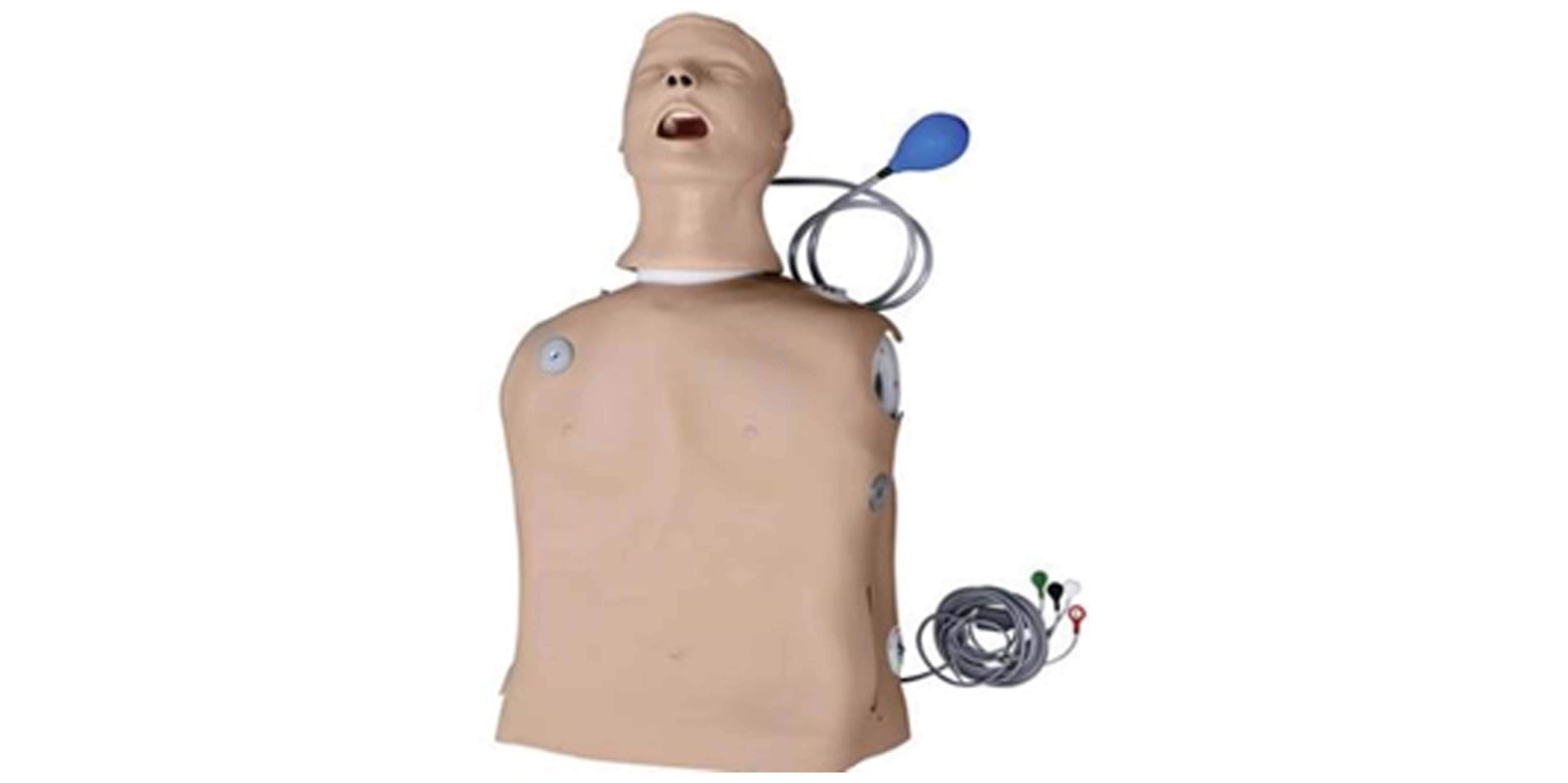 |
FR2 AED Teaching ModelTen pre-configured sudden cardiac
arrest scenarios compatible with training programs developed by
internationally recognized responder programs. Remote
control (optional) for manual scenario selection pause/resume function,
AED Trainer 2 volume control, motion artifact, low battery or replace
battery, loose electrodes connection, shockable or nonshockable rhythms,
and error conditions that simulate the red X in the HeartStart FR2’s
status display window.
Pull-tab status indicator simulates the
Heartstart FR2 status window for maintenance training. It includes high-quality soft pack with “1-2-3” protocol card for added training realism and durable storage of the equipment. | |